Introduction
The egg polarization index or PI provides a very good indication of female readiness to spawn, but by itself it does not measure directly the quality of the eggs. A biological test or egg maturation assay can be performed to determine if the eggs are in the stage of final maturation and will likely respond to hormonal injections. Immature eggs may be induced to ovulate but they cannot be successfully fertilized until they complete final egg maturation. The capacity of the egg to undergo final maturation can be adversely affected by the animal's own physiological or metabolic response to unfavorable environmental or husbandry conditions such as abrupt temperature changes and rough or frequent handling. Eggs can also be overripe, a condition of degeneration or reabsorption of yolk nutrients (atresia). Like immature eggs, overripe eggs can also be induced to ovulate but are often of poor quality. Overripe eggs often correlate to low fertility or a high percentage of embryo and larval deformities, accompanied with low survival.
Principle
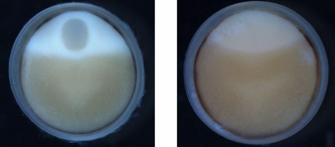
During the period of yolk accumulation, the eggs of sturgeon are in an arrested stage of cell and nuclear division (i.e., meiosis is arrested) and are considered immature; even though the female may have 'black-eggs'. Close to ovulation, when the female is ready to spawn, the eggs reach a species-specific determined size and the eggs are ready to resume meiosis and enter the final stage of egg maturation. During the stages of final egg maturation, the nucleus or germinal vesicle (GV) begins to migrate to the edge of the egg where it undergoes germinal vesicle breakdown or GVBD. Final egg maturation is accompanied by cytoplasmic maturation which is clearly observable in the segregation and polarization of the different size yolk granules within the egg cytoplasm. Once GVBD occurs, the egg is released or ovulated from the follicle. Once released the egg is now referred to as an ovum and the genetic material of the female, contained within the now broken egg nucleus, can mix with the genetic material of the male, carried inside the egg cytoplasm by the fertilizing sperm.
The process of final egg maturation is under control of a maturation inducing-substance or MIS. Progesterone and several of its derivatives are considered the most potent hormones to induce final egg maturation in many animals, including sturgeon. The process of final egg maturation and GVBD can be induced artificially by in-vitro incubation of eggs in a solution of progesterone.
Spawning success and viability of the eggs are primarily influenced by the size of the egg, PI, and stage of egg maturation. A sample of eggs of the right size (i.e., species-specific determined egg diameter), a low PI (less than 0.10), with distinct yolk polarization, and 100% GVBD can be reliable indicators of a female sturgeon's readiness to spawn and the viability of her eggs.
Materials
- For fish handling: tube-net, stretcher and sawhorses, freshwater source.
- For gonad biopsy: surgical gloves, catheter of flexible tubing at least 4 mm ID or modified trocar, #3 scalpel handle and #10 or #15 blades, Adson-Brown tissue forceps 7x7 teeth, Olsen-Hegar needle holder scissors combination, absorbable sutures (e.g., Vicryl-polyglactin 910, size 0 or 1 with OS-6 or 4 cutting needle; PDS II-polydioxanone, size 1 with CP-1 cutting needle), 1% iodine solution, sterile wipes, alcohol or iodine for cleaning/sterilizing instruments in between samplings, and a small "sharps" container for the used blades and suture needles.
- For the egg samples: 25 mL plastic vials with caps or 50 mL centrifuge tubes or tissue culture flasks, culture medium (e.g., Ringer solution or Leibovitz L-15, in 1 L plastic bottles), two holding coolers with gel paks or wet ice, to maintain medium and to transport the containers of eggs at 14–18°C, balance (or access to one) and accessories to weigh chemicals (NaCl 6.5 g, NaHCO3 2 g, CaCl2 300 mg, and KCl 250 mg) to make Ringer solution when added to 1 L of distilled or de-ionized water.
- For the progesterone hormone assay: disposable plastic transfer pipettes (to handle/transfer eggs), 30 mL glass beakers, tissue culture plate/vessel (e.g., 4 multi-well, or petri dishes single or with 3 partitions, or 100 mL beakers or 125 mL Erlenmeyer or 150 mL fleaker flasks), progesterone hormone stock solution of 2 mg progesterone/1 mL ETOH: weigh 10 mg progesterone (Sigma P-8783) and dissolve in 5 mL ethanol/ethyl alcohol (absolute or 95%); or use water-soluble progesterone (Sigma P-7556, that contains approx. 7 mg of progesterone in 100 mg of powder chemical). Weigh 20 mg of water-soluble progesterone and dissolve in 1.4 mL distilled water for a stock solution of 1 mg water-soluble progesterone in water. The alcohol solution can be stored for several weeks.
- For assay analysis: disposable plastic transfer pipettes (to handle/transfer eggs), 20 mL glass beakers, boiling stones, aluminum foil, hot plate, oven mitts, timer, 25 mL plastic vials with caps, 10% formalin fixative (9 parts water: 1 part formaldehyde), tools to handle and cut the eggs: delicate Adson-Brown forceps, double-edge razor blades (these are the thinnest), cutting surface (e.g., Petri dish bottom), ice tray/bucket, illuminated desk magnifier or dissecting/stereo/macro-scope.
- Miscellaneous: data sheets or notebook, pens/pencils, general-purpose gloves, wipes, and paper towels.
Protocol
Females are sampled during their expected month of spawning (see Sturgeon Aquaculture: Specialized Techniques: The Egg Polarization Index or PI, for procedures). Sampled eggs are analyzed for PI and also tested for their capacity to undergo GVBD in the presence of progesterone. The in-vitro egg maturation assay is conducted in a modified Ringer solution or L-15 at 16°C for at least 16 hours. Fifteen to twenty eggs are incubated in a solution containing 5 µg/mL progesterone and without (controls). After incubation, the eggs are boiled, chilled on wet ice, stored in 10% buffered formalin, bisected, and examined for GVBD.
- Add 20 mL of Ringer solution to each clean egg incubation vessel, or adjust the volume (2x, 3x, etc) depending on the size of the incubation vessel (the eggs need to be covered with solution). Set up two containers for each female (control and progesterone). Label them C (control) or P (progesterone), and include the brood fish identification number, if you are setting up assays for several different females at one time.
- Transfer 15–20 eggs into another clean container (e.g., small beaker or dish), using a clean (unused) disposable plastic pipette. Carefully remove all solution that was also transferred to the beaker, by pouring it off or pipeting it out. Then pipette into the beaker, about 5 mL of Ringer's from the dish or flask the eggs will go into, gently swirl the beaker and pour the eggs and media back into that container (this is an important step to maintain the correct volume of Ringer solution in each dish, well or flask).
- Add either 50 uL or 200 uL of the progesterone stock solution (i.e., 2 mg progesterone/mL alcohol or 1 mg water-soluble progesterone/mL in water stock solutions, see Materials) with a micropipette or 1 cc syringe, to the progesterone labeled containers (or adjust the amount based upon the amount of solution the eggs are in). The control containers should receive the same quantity (50 µL or 200 uL) of the carrier (95% ethanol or water) used to dissolve the progesterone. To avoid any cross-contamination, use separate syringes for progesterone and controls, and if a micropipette is used, change the tip. Gently swirl the dishes or flask to mix the solutions. Be extremely careful in preventing contamination of controls with progesterone, because even minor exposure to progesterone may cause GVBD in the controls. Place the control plates on the top shelf of a temperature-controlled incubator that is accurate to ± 0.5°C (see Materials), and keep the progesterone containers on the bottom, to prevent any potential spills from the top to the bottom shelf that may affect your results.
- Record the time and incubate for 16–18 hours at 16°C. If the assays are set-up at 4 pm they will be finished at 8 am the next morning. Make sure the incubation cabinet is set at 16 ± 0.5°C. It should be turned on and running at this temperature for one week prior to the time of sampling.
- When incubation is completed, using pipettes (one labeled control and one progesterone) transfer the eggs to individual 20 mL beakers, labeled with the female identification number and a C (control) or P (progesterone). Add to each beaker Ringer stock solution to reach a total volume of 15 mL and boil gently for 1–3 minutes. An equal volume in all beakers will ensure that they all boil at similar rates. Add a boiling stone to each beaker and cover them all with a sheet of aluminum foil, as the eggs will occasionally "pop-out" of the beaker during boiling.
- Immediately cool the eggs by placing the beakers directly on crushed ice for 15–30 minutes.
- At this time the eggs could be cut with a razor blade and evaluated. However, placing them in 10% buffered formalin at least for 30 minutes or overnight will make cutting much easier. Very mature females usually have soft eggs, even after chilling, but they become firmer and easier to cut after storage in buffered formalin.
- Section each egg along the animal-vegetal axis, usually recognized by the oval shape of the freshly collected egg. Turn both halves section side up, and observe the presence or absence of the germinal vesicle by focusing a light beam on the section surface. Record germinal vesicle breakdown (GVBD) or intact germinal vesicle for each egg (Figure 1). Eggs from both control and progesterone solutions should be examined, making notes on germinal vesicle shape, size, and whether it migrated to the very top of the animal pole. If you are not sure you properly sectioned the egg, take the two halves and cut them again (into quarters); this will undoubtedly reveal any GV that may have been missed in the first cut.
- Evaluate the results by calculating the percentage of eggs that underwent GVBD in the control and progesterone treatments. A 100% response in the progesterone and some response in the controls indicates that you should spawn the female within 1 week. A 100% response in the progesterone and no response in the controls indicate that you should probably spawn this fish in about 2 weeks. A response of <100% in the progesterone indicate that you should re-sample in approximately 3–4 weeks. These time intervals are assuming the females are being held at approximately 13°C–15°C. At colder temperatures these time periods maybe slightly extended and at warmer temperatures the time periods should be reduced.

Credit: UF/IFAS