Summary
- Petiole (rachis) blight is caused by numerous fungal pathogens, but the symptoms these pathogens cause are similar for all of them.
- Palm host range is unknown for this disease.
- The disease often results in discolored (usually brown or reddish-brown) elongated lesions or streaks along the petiole and/or rachis of the oldest (lowest) leaves.
- The pathogens infect only the petiole or rachis, not the leaf tissue. However, destruction of vascular tissue (xylem and phloem) deep into the petiole results in a one-sided or uneven death in the leaf blade.
- Fungal structures can sometimes be observed on the infected petiole surface.
- Since very little is known about this disease, only general disease management strategies regarding sanitation, water management, and fungicide use can be provided.
Introduction
To discuss petiole (rachis) blight, a brief introduction to palm leaf anatomy is required. There are three types of palm leaves. All possess a base (leaf sheath), a petiole, and a blade. The leaf base is the portion that attaches to the stem (trunk). The leaf petiole is the stalk or stem of the leaf connecting the leaf base with the leaf blade.
The leaf blade is either: 1) fan-like (palmate) with individual leaflets (segments) merged together and the petiole terminated where the blade begins; 2) an elongated version of fan-like (costapalmate) with individual leaflets (segments) merged together but with the petiole extending into the leaf blade; or 3) feather-like (pinnate) with individual leaflets clearly delineated and attached to the petiole, with the petiole extending the entire length of the leaf blade. The extension of the petiole into the leaf blade is called the costa on a costapalmate palm, and the rachis on a pinnate palm.
Unfortunately, the term petiole and rachis are often used interchangeably. Since diseases of the petiole were first observed on Phoenix palms, pinnate-leaf palms that have a rachis, the common name of these diseases became rachis blight and it is not uncommon to see the disease on both the petiole and the rachis. However, as we now know, palms that do not have a rachis also are susceptible to petiole diseases. In this fact sheet the disease will be referred to as petiole blight.
In general, the typical or classic petiole blight is a disease in which the pathogen only infects the petiole and rachis. The disease symptoms may look like the pathogen has also infected the leaf blade, but this is a secondary affect from petiole infection. An exception to the typical petiole blight is a disease caused by Pestalotiopsis. This pathogen causes both a leaf spot and blight as well as a petiole or rachis blight, usually at the same time. It is discussed in a separate fact sheet at https://edis.ifas.ufl.edu/pp141.
Pathogens and Hosts
There are a number of different pathogens associated with petiole blight. These include Neodeightonia, Cocoicola, and Serenomyces, which are only found in association with palms. Macrophoma, Phoma, Phomopsis, Diplodia, and Lasiodiplodia may also cause this disease, but these fungi also cause diseases of numerous other plant hosts. The host range of all of these fungi within the palm family is unknown.
Symptoms
Mature palms (palms with trunks) are more likely to be affected by petiole blight than juvenile palms. In both juvenile and mature palms, the oldest (lowest) living leaves will be affected first. The disease will move upward through the canopy to younger leaves. The disease may kill a number of the lowest leaves, but the disease seldom kills the palm (Figure 1).
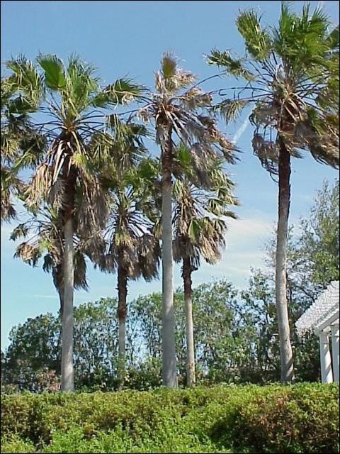
Credit: A. Wilson
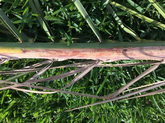
No matter which pathogen is causing the disease, the symptoms of petiole blight are essentially the same. Discolored (usually brown or reddish-brown) elongated lesions or streaks often occur along the petiole and rachis (Figure 2). The fungus invades deep into the petiole and destroys all tissue, including the vascular tissue (xylem and phloem). This vascular tissue moves water and carbohydrates between the leaf and the stem (trunk). Vascular tissue destroyed in the petiole or rachis results in localized death in the leaf blade because only the leaf segments or leaflets connected to the destroyed vascular tissue are killed (Figure 3). This results in a one-sided or uneven death of the leaf blade, the symptom most often observed first. For Phoenix species, Syagrus romanzoffiana (queen palm), and Washingtonia species, these symptoms appear similar to those caused by Fusarium wilt, which is a lethal disease. Only a laboratory diagnosis can differentiate the cause of the symptoms observed in these three palm genera. See the fact sheet on these diseases at https://edis.ifas.ufl.edu/pp139 and https://edis.ifas.ufl.edu/pp278.
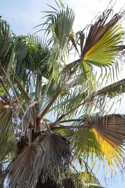
Credit: M. L. Elliott
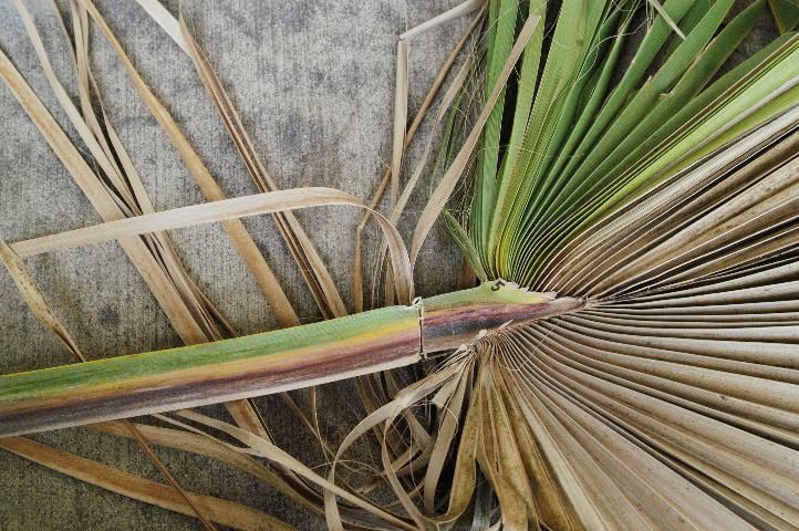
Credit: M. L. Elliott
Again, while it appears the fungus is also infecting the leaf blade, it does not. The leaf blade symptoms are a result of the fungal damage in the petiole and rachis. Thus, for diagnostic purposes, the petiole or rachis is the tissue that must be examined and sampled, and not the leaf blade. A cross-section through the petiole or rachis lesion or streak will reveal the discolored internal tissue resulting from the fungal invasion. It is critical to use a saw to cut through the petiole or rachis. A tool such as a pruning shear may bruise internal, healthy plant tissue in the cutting process. This bruise can then be mistaken for fungal damage.
Sometimes, the surface of the diseased petiole or rachis will have erupted with fungal structures while the leaf is still attached to the palm trunk (Figure 4). These structures either are or can be induced to produce spores used to identify the pathogen.
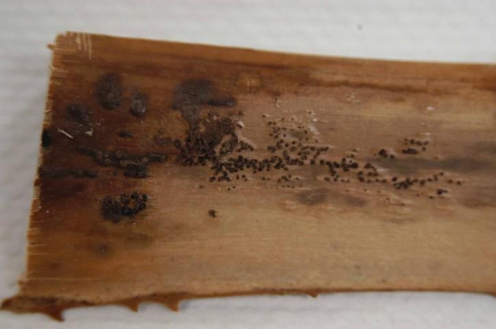
Credit: M. L. Elliott
Diagnosis
As stated above, fungal structures may be present on the petiole or rachis at the time the symptoms are observed. These structures may also be sporulating while the leaf is still attached to the trunk, which makes pathogen identification relatively simple. If not producing spores, it is usually possible to induce spore production. If these structures are not immediately evident, placement of the diseased petiole or rachis segment into a moist chamber (e.g., sealed plastic container with wet paper toweling) will often induce their development (Figure 5).
Credit: C. Hass
While some of the pathogens that cause petiole blight can be cultured on artificial media, many cannot. The Florida Extension Plant Disease Clinic (FEPDC) network is available for pathogen identification. Contact your local UF/IFAS Extension office or FEPDC for information on sample submission and cost of laboratory diagnosis. At a minimum, a 2-foot section of the petiole or rachis exhibiting the typical symptoms should be submitted for diagnostic purposes. The section can be cut into pieces for easier packaging. Do not place the pieces in a plastic bag. Simply place in a cardboard box and pad with newspaper.
Disease Management
Very little is known about petiole blight, including the specific environmental conditions that encourage disease development. It is presumed that high humidity is required for fungal infection and disease development, as that is a requirement for many plant diseases that affect above-ground tissue. It is also presumed that spores are the primary means of fungal movement from palm to palm. Based on these assumptions, sanitation and water management are the basis for petiole blight management.
Removal and destruction of severely infected leaf fronds would be suggested as a means of reducing inoculum (available spores to infect other leaves and palms). This would be especially critical in a nursery situation where palms are planted closer together and are more numerous. However, if the palms are in the landscape and under nutrient stress, pruning should be minimal as removal of too many leaves may be detrimental to the palm rather than beneficial.
In a nursery, increasing distance between plants, increasing air movement, and irrigating in the early morning hours to reduce leaf wetness at night are critical water management components. When possible, overhead irrigation should be eliminated.
Since no fungicides have been evaluated for efficacy in managing petiole blight, fungicide recommendations are educated guesses at best. Fungicides would only be recommended in those situations where a palm is being seriously affected, or the potential for an epidemic is high, such as in a nursery. Never initiate a fungicide program without first initiating a cultural control program as described above.
For palms where the canopy is still accessible for spraying, broad-spectrum contact or systemic foliar fungicide sprays to protect the petiole and rachis may be useful. A fungicide that will adhere to the waxy petiole or rachis tissue would be most useful.
It is critical to understand that fungicides do not cure the damage already present on the palm. Plant tissue does not heal itself. Once the damage occurs, it will remain for the duration of the life of that particular leaf. Fungicides are used to prevent further spread of the disease by protecting a leaf petiole and rachis that has not yet been infected by the fungal pathogen.
Selected References
Barr, M. E., H. D. Ohr, and M. K. Murphy. 1989. "The genus Serenomyces on palms." Mycologia 81: 47–51.
Elliott, M. L. and E. A. Des Jardin. 2006. "First report of a Serenomyces sp. from Copernicia x burretiana, Latania loddigesii, and Phoenix canariensis in Florida and the United States." Plant Health Progress. doi:10.1094/PHP-2006-1213-02-BR.
Elliott, M. L. and E. A. Des Jardin. 2006. "First report of Cocoicola californica on Washingtonia robusta in Florida." Plant Health Progress. doi:10.1094/PHP-2006-0227-01-BR.
Elliott, M. L. and E. A. Des Jardin. 2014. Serenomyces associated with palms in southeastern USA: isolation, culture storage and genetic variation. Mycologia 106:698-707. doi:10.3852/13-353.
Hyde, K. D., and P. F. Cannon. 1999. Fungi Causing Tar Spots on Palms (Mycologial Papers). Wallingford, U.K.: CABI Publishing.