Introduction
Phorid flies (Diptera), also known as humpback flies or scuttle flies for their appearance and behavior, are an extremely diverse group of flies that are saprophagous (feed on decaying organic matter), parasitic, or phytophagous (feed on plants). Within the Phoridae family, the genus Megaselia is also extremely diverse, with more than 1400 described species, many very similar in appearance. The name "cob fly" was given to a Megaselia spp. that attacked corn in Texas (Walter and Wene 1951). To date, only one described species (Megaselia seticauda Malloch) has been observed to damage sweet corn. This species was originally identified as the ubiquitous saprophagous fly Megaselia scalaris, but was later correctly identified as M. seticauda (CDFA 1996).
Geographical Distribution
Megaselia seticauda was first collected in Costa Rica from an area cleared for pasture, corn, and potatoes (Cresson and Malloch 1914). There are reports of damage to immature "green corn" ears from Texas (1944), Mexico (1942), and Ecuador (1954). It has been collected in Brazil (1962), and it was detected in California in 1996 (Walter and Wene 1951, Borgmeier 1962, CDFA 1996).
In Texas, M. seticauda was found in a few corn ears in 1944, but by 1950, fields that were not treated with DDT for corn earworm were severely infested with both M. seticauda and the silk fly Euxesta stigmatias (Diptera: Ulidiidae). Larvae of both E. stigmatias and M. seticauda were reported as being relatively unaffected by DDT spray residues, leading to the recommendation to treat adults (Walter and Wene 1951).
In Florida, Megaselia spp. flies, possibly M. seticauda, have been found since the late 2000s in sweet and field corn ears that were not protected by insecticides and were grown during the spring and summer. These phorid larvae feed on silks, cobs, and kernels as do silk flies, but phorid larvae develop faster and cause damage quicker than silk fly. When observed in a field immediately before silking, these phorids should be regarded as an economic concern, and this document was written with the intent of helping crop consultants identify this fly. The following information is based on our field and laboratory observations of live immature and adult stages of this fly species. There are very little data on the economics, population dynamics, seasonality, or insecticide susceptibility of corn-infesting Megaselia spp. More research is needed to improve our understanding of how this fly impacts Florida sweet corn production.
Distinguishing Cob Fly from Similar Corn Ear Pests
Egg
Dozens of cob fly eggs are deposited in the silks at tip of corn husks. There have been reports of fly eggs deposited on husk leaves before silks emerge; these may be either cob fly or silk fly eggs.
Larvae
In ears of corn, larvae of phorids can be distinguished from larvae of silk flies by the tapering shape of the posterior abdomen (Figure 1). In contrast, silk fly larval abdomens terminate bluntly, and two dark-colored, peg-like spiracular (breathing tube opening) plates can be easily seen at the end of the abdomen (https://edis.ifas.ufl.edu/in381). These spiracular plates are not present in cob fly larvae. Phorids retain their white coloration throughout the larval stage, while silk flies darken to a yellow color in the third instar. When disturbed, silk fly larvae will grab their spiracular plates with their mouth hooks on the head, flex, and jump several centimeters. Phorid larvae do not exhibit this behavior.
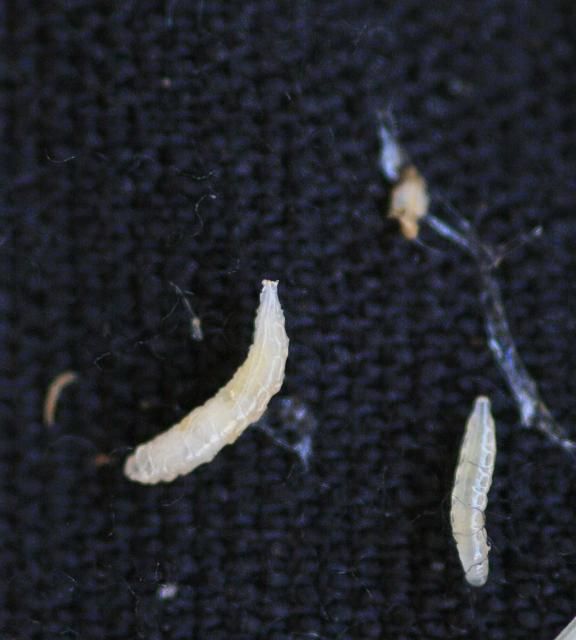
Credit: D. Owens, UF/IFAS Everglades Research and Education Center, Belle Glade, FL
When the larvae emerge from the eggs, they quickly enter the ear to feed on silks, cob, and kernels. Cob fly larvae can be observed as a larval "mass" that feeds through the silks and destroys them before reaching the ear tip (Figures 2 and 3). Larvae develop and damage silks quickly enough to interfere with pollination. Once they reach the ear, larvae feed on the cob and developing kernels at the tip (Figures 4 and 5). Cob flies leave the ear 10–14 days after first silk, usually before blister stage, by leaving a visible shiny mucous trail on the drying silks (Figure 6). For comparison, silk fly larvae are still developing in the ear until the ear is in milk stage and ready for harvest. Silk flies require 12–19 days from oviposition to mature larvae leaving the ear. Silk flies also do not leave a mucous trail when they exit ears. When cob fly populations are high, damage may also be observed on ears and tassels of plants that developed as suckers from the main plant, as well as on secondary and tertiary ears beneath the primary ear on the main shoots.
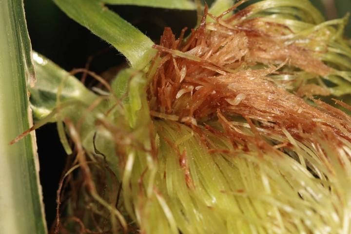
Credit: D. Owens, UF/IFAS Everglades Research and Education Center, Belle Glade, FL
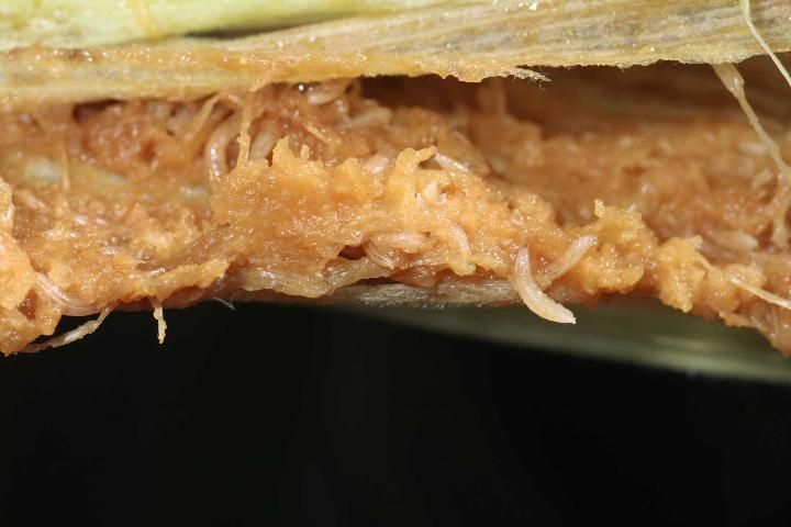
Credit: D. Owens, UF/IFAS Everglades Research and Education Center, Belle Glade, FL
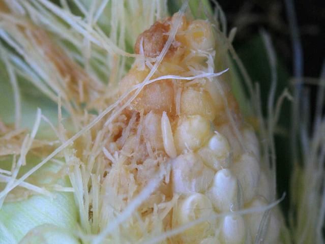
Credit: D. Owens, UF/IFAS Everglades Research and Education Center, Belle Glade, FL
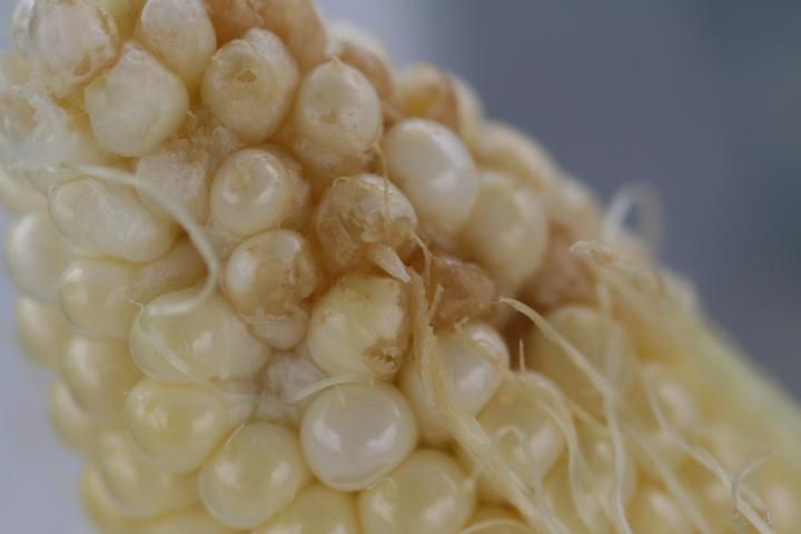
Credit: G. S. Nuessly, UF/IFAS Everglades Research and Education Center, Belle Glade, FL
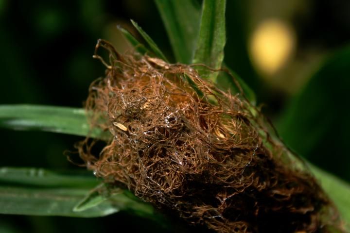
Credit: G. S. Nuessly, UF/IFAS Everglades Research and Education Center, Belle Glade, FL
Pupae
Most cob fly larvae leave the ear at the end of larval development (end of the 3rd instar) and pupate in soil. Occasionally, pupae can be found in the dried silks or rarely between kernels in the ear. Pupae are light brown, somewhat triangular and noticeably flattened, with ridges across the surface. Two narrow, black respiratory horns are prominently displayed on the posterior end (Figure 7). Silk fly pupae, which are reddish brown, elongate, cylindrical, and only slightly flattened with the anterior end tapered to a blunt point, are easily separated from these phorid pupae. Silk fly pupae have large black oval spiracles at the posterior end. We do not currently know how long pupal development requires in the field.
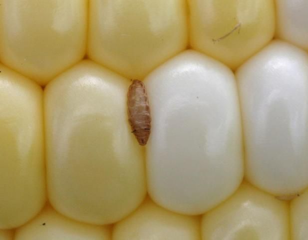
Credit: D. Owens, UF/IFAS Everglades Research and Education Center, Belle Glade, FL
Adult
Adult cob flies are 2–3 mm in length, about half the length of silk flies (4–7 mm). Adult phorids are light brown with dark brown to black markings on the dorsal side of the abdomen. Hind femurs have a dark pigmentation at the end. These corn-feeding phorids have no dark bands across their wings, further distinguishing themselves from silk flies. Phorid wing venation is very distinct, with heavy veins curving to meet the wing margin about halfway between the wing tip and the body. Smaller, lighter veins radiate out from the thick veins. The head is small and has large, black bristles (Figures 8 and 9). Phorid flies have a very distinctive locomotion. They move very quickly in a "herky-jerky" fashion characterized by short, rapid running, a pause, a turn, and more short, rapid running.
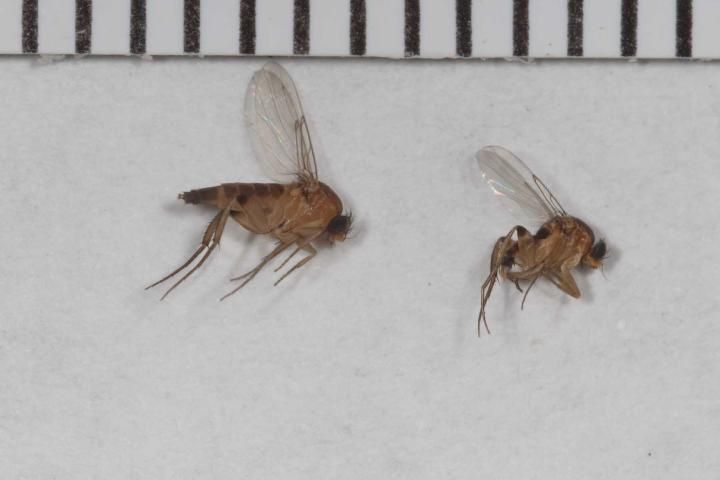
Credit: D. Owens, UF/IFAS Everglades Research and Education Center, Belle Glade, FL
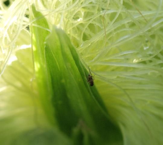
Credit: D. Owens, UF/IFAS Everglades Research and Education Center, Belle Glade, FL
Damage
Cob fly damage to ears may be widespread less than 7 days after first silk. Damaged silk tissue is reddish brown to brown (Figure 10). Heavily damaged silk appears wet and slimy and may be a site of secondary infection by pathogens. Severe tip damage can occur on ears that are still at least one week to ten days from harvest (Figure 11). Field corn ears infested with phorid larvae frequently display pollination disruption with the top 25 to 50% of the cob bare of kernels at harvest (Figure 12). Because larvae develop faster and leave the ear earlier than silk fly, it is possible that phorids are partly responsible for the damage often attributed to silk fly.
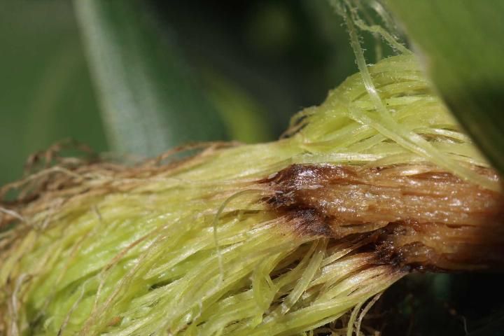
Credit: D. Owens, UF/IFAS Everglades Research and Education Center, Belle Glade, FL
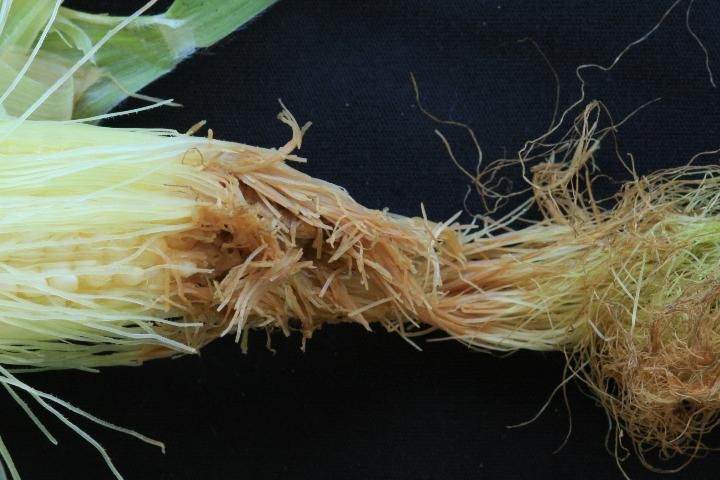
Credit: D. Owens, UF/IFAS Everglades Research and Education Center, Belle Glade, FL
Management
Scouting corn for cob flies should begin at tassel-push (as the tassel emerges from the plant at the beginning of the reproductive stage). Treatment may be justified prior to first silk or as early as ear shoot emergence, if phorids adults are present. Experimental fields at UF/IFAS Everglades Research and Education Center in Belle Glade, FL that received multiple insecticide applications per week for silk fly control exhibit reduced cob fly damage. Therefore, insecticides used for silk fly management likely provide cob fly control.
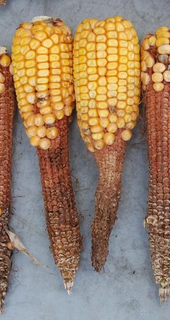
Credit: G. S. Nuessly, UF/IFAS Everglades Research and Education Center, Belle Glade, FL
References
Borgmeier, T. 1962. "Versuch einer Uebersicht euber die neotropischen Megaselia-Arten, sowie neue oder wenig bekannte Phoriden verschiedener Gattungen (Dipt. Phoridae)." Studia Ent. Petropolis 5: 289–488.
California Department of Food and Agriculture (CDFA). 1996. California plant pest and disease report, v 15: 150–181. Ed. R. J. Gill. https://www.cdfa.ca.gov/plant/ppd/PDF/CPPDR_1996_15_5-6.pdf. Verified 22 April 2016.
Cresson, E. T., Jr., and J. R. Malloch. 1914. Costa Rican Diptera collected by Philip P. Calvert, Ph.D., 1909–1910. "A partial report on the Borboridae, Phoridae, and Agromyzidae." Trans. Am. Entomol. Soc. 40: 1–36.
Walter, E. V., and G. P. Wene. 1951. "Tests of insecticides to control larvae of Euxesta stigmatias and Megaselia scalaris." J. Econ. Entomol. 44: 998–999.