This paper presents an overview of micropropagation to help growers understand and evaluate basic plant tissue culture propagation for their own nursery operations. Micropropagation is a form of tissue culture used to rapidly propagate or regenerate a large number of plants.
Introduction
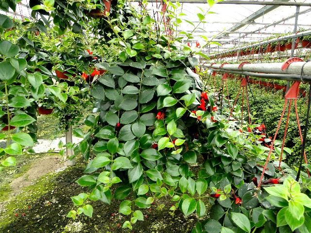
Florida leads the nation in foliage plant production, accounting for $454 million of the $631 million national wholesale value in 2013 (USDA 2014). Many nurseries propagate their own liner materials by cutting, dividing, grafting, or air layering from stock plants maintained on their premises (Figure 1). Growers must calculate the costs per square foot of in-house propagation, which includes the cultural costs of maintaining the stock, labor costs of maintenance and propagation, and revenues per square foot of greenhouse space assigned for stock not generating the direct income production footage provides.

Credit: J. Chen, UF/IFAS
Over half of all tropical foliage crops are now propagated via micropropagation methods (Table 1). Micropropagation is cost effective because large numbers of disease-free plantlets can be originated in a tissue culture laboratory. Micropropagation dramatically reduces greenhouse space required for maintaining stock plants, helps eliminate systemic diseases in starting materials, and provides growers with healthy, uniform liners year-round.
Development of Micropropagation in Horticulture
The foliage plant industry was the first to successfully demonstrate the commercial profitability of micropropagation. Applying micropropagation to tropical foliage plants began in the late 1960s with the development of commercial laboratories (Chen and Henny 2008). As one of the major technological advances in the global horticultural marketplace, micropropagation now annually produces worldwide more than 507 million ornamental tropical foliage plantlets, including more than 150 million orchids (Chen and Stamps 2006).
In 1974,Toshio Murashige, an early researcher in the field of tissue culture, defined three stages of micropropagation that later grew to four stages: Stage 1, establishment of clean cultures; Stage 2, multiplication; Stage 3, root initiation; and Stage 4, acclimatization. Stage 3 and Stage 4 have been blended in commercial practice (Chen and Henny 2008). Recently, a Stage 0 has been added. Stage 0 is the maintenance of stock plants under clean nursery conditions prior to selecting them for tissue cluture Stage 1.
Stage 1. Initiating Plant Tissues into Test Tube Culture (from In Vivo to In Vitro)
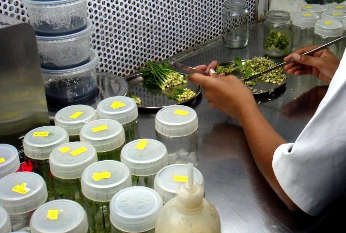
Shoot and root tips, stem nodes, axillary buds, and flower parts may all be harvested for micropropagation. Plant tissues are excised or removed from original stock and sterilized, usually with a bleach or alcohol wash. These tissues, at this point referred to as explants, are placed into the aseptic environment of a test tube, petri dish, or culture vessel where a sterile agar or gel-based culture medium enriched with nutrients, sugars, vitamins, minerals, and plant hormones induces rapid tissue multiplication. This stage of tissue culture is the most costly, particularly when new plant genera or new varieties are initiated into culture.

Credit: J. Chen, UF/IFAS
Stage 2: Multiplication
If the explant was not sterilized completely during Stage 1, bacterial or fungal growth will be evident within days. Bacterial or fungal growth will have destroyed the cultures, and Stage 1 will need to be repeated. If the cultures are clean, the explant will produce noticeable cell growth within about four weeks. Under the correct nutrient and hormone recipes, explant tissue will multiply and begin to grow new shoots.
If the new shoots were derived from explants of terminal or lateral meristems, the shoots are termed axillary shoots. Axillary shoots reliably produce true-to-type plantlets because they grow from pre-existing meristems. Axillary shoots are identical to the original explant and can be subcultured indefinitely.
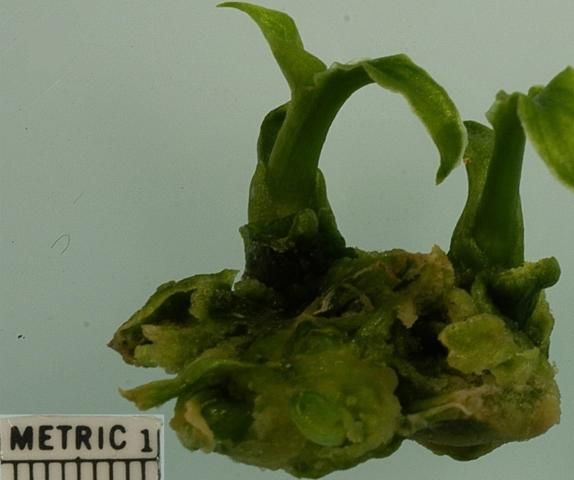
If the new shoots were derived from tissues other than meristems, the shoots are termed adventitious shoots. These tissues might be leaf tissue, petiole tissue, stem tissue, root tissue, or flower parts, such as ovules. Adventitious shoots can produce plantlets that are not true to type. In tissue culture, these non-meristematic parts give rise to undifferentiated cells called "callus." Callus can be induced to develop adventitious shoots with the proper hormone and nutrient regimen, however; propagation through the callus phase sometimes produces variation in subsequent plantlets. This is termed somaclonal variation. To avoid such variation, commercial tissue laboratories normally propagate foliage plants from existing meristems (Figure 3).

Credit: J. Chen, UF/IFAS
Stage 3: Root Initiation
Traditionally, Stage 2 shoots were transferred to a gel or agar medium containing auxins, a class of plant hormones that induce root development. Before the plantlets were transplanted into a soilless potting mix (Stage 4), plantlets remained in laboratory culture until roots developed (Stage 3). In current commercial practice, unrooted shoots (microcuttings) produced in Stage 2 are transferred from culture directly to a potting mix, where natural levels of auxins are sufficient to initiate rooting.
Stage 4: Acclimatizing Tissue-cultured Plants
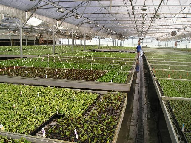
Tissue-cultured plants are grown under uniform laboratory conditions. Greenhouse conditions must be well controlled when microcuttings are transferred into the potting mix (Hennen 2003). There are two significant challenges to overcome in acclimatization: poor control of water relations and decreased photosynthetic competence in the plants produced in vitro. The tissue culture laboratory may provide acclimatization, or the nursery operator may purchase unrooted microcuttings and perform the acclimatization stage at the nursery (Figure 4).

Credit: Richard J. Henny, UF/IFAS
Microcuttings should be given immediate attention when they arrive at the nursery. A grower should avoid temperature extremes on the shipping dock. Growers should provide clean tools and trays for the arriving plantlets.
Microcuttings are usually potted into cell trays filled with premoistened peat mix. The pH in the potting mix should be adjusted to between 5.8 and 6.1. Total soluble salts should initially be kept low, and liquid fertilization may be supplied at 100 to 150 ppm.
Because light levels in the tissue culture laboratory are low, commonly 200–600 f.c. (2.2–6.5 klux), growers would want initial light levels for the microcuttings also kept low at 1,800–2,000 f.c. (19–22 klux). Light levels can gradually be increased to 1,800 –2,500 f.c. (19–27 klux). During the hottest, brightest part of the day, growers might want to keep newly potted plantlets shaded with cheesecloth or shadecloth. The light levels may be doubled every two weeks until plants are acclimated.
A mist or fog system is suggested to prevent desiccation. Growers can gradually increase mist intervals until plants adapt. It is advised to monitor the plants closely and treat fungal diseases immediately to prevent losses.
Advantages of Micropropagation
Micropropagation helps eliminate systemic diseases in starting materials, dramatically reduces greenhouse space required for maintaining stock plants, and provides growers with healthy and uniform liners year-round.
To be considered truly disease-free, plants from micropropagation may require using phytopathological disease eradication techniques such as meristem or meristem-tip culture. In more difficult cases, chemo- or thermotherapy may be needed to eradicate a disease agent from the plant tissue.
Micropropagation has allowed propagators to select desirable variants and generate new, viable cultivars more quickly and easily; more than 80 commercial cultivars have been selected from variants and introduced into the trade.
Micropropagation also speeds the introduction of hybrid cultivars. Through micropropagation, new cultivars reach sufficient numbers to become commercially available in 2 to 3 years, whereas cultivars produced with traditional propagation methods require 5 to 7 years.
Nursery operators and growers who want to further investigate micropropagation should visit one of the many horticultural trade shows to see commercial tissue culture propagators display their microcuttings. At trade shows, growers can view the microcutting products, obtain detailed information regarding their specific crops, and evaluate costs.
References
Chen, Jianjun, and Richard J. Henny. 2008."Role of Micropropagation in the Development of the Foliage Plant Industry." In Floriculture, Ornamental and Plant Biotechnology Volume V. 206–218. UK: Global Science Books Ltd.
Chen, J., and R.H. Stamps. 2008. "Cutting Propagation of Foliage Plants," In Cutting Propagation: A Guide to Propagating and Producing Floriculture Crops, edited by J.M. Dole and J.L. Gibson , 203–28. Batavia, IL: Ball Publishing.
Hennen, Gary. 2003. "Acclimatizing Tissue-Cultured Plantlets," In Ball RedBook Volume 2 Crop Production, edited by D. Hamrick, 172. Batavia, IL: Ball Publishing.
USDA (United States Department of Agriculture). 2014. Floriculture Crops 2013 Summary. Washington, DC: USDA.