Abstract
This article is meant to serve as a general guide to perform quantitative fecal egg counts on grazing small ruminants, specifically sheep and goats. This tool is utilized to estimate the extent of pasture parasite burden as well as individual animal parasite burden, and to determine the efficacy of dewormer or anthelmintic treatment. Producers should discuss interpretation of fecal egg counts and treatment decisions with their veterinarians, because a valid veterinarian-client-patient relationship (VCPR) is legally required to make such recommendations.
Introduction
The most common test performed to evaluate quantitative fecal egg counts on grazing animals is the modified McMaster’s test or McMaster’s fecal egg count (FEC). This procedure is relatively inexpensive and noninvasive. Producers have the option to send animal fecal samples to a commercial laboratory for analysis or, with the proper tools and training, to analyze samples themselves as detailed in this publication. A fecal flotation is the best method to screen for overt parasites in a given species, but it cannot be used for quantification. Numerous parasites can inhabit their host’s gastrointestinal tract to produce eggs, larvae, or cysts that ultimately exit the host’s body via feces. McMaster’s FEC can be used as a tool to aid in diagnostic decision-making for anthelmintic treatment purposes. The FEC is not an absolute representation of parasite burden, as many factors influence egg production in the host such as host immunity, nutrition, stress, concurrent infections, age, pregnancy status, and more.
Integration with Other Assessment Techniques
While fecal egg counts are a valuable tool for measuring potential parasite burden and anthelmintic efficacy, they should not be used in isolation. Fecal egg counts do not correlate to actual worm numbers or to the severity of parasitic disease. Some species of worms lay more eggs than others, and some are more dangerous than others. Integrating FAMACHA® scoring and other visual assessment techniques, such as the Five Point Check©, is recommended to supplement FEC data for effective parasite management. These methods help to assess the overall health of the animals and to identify those needing treatment, determinations which in turn reduce the use of dewormers and help to prevent the development of anthelmintic-resistant parasites.
Collection of Fecal Samples
Fecal evaluation should be conducted on fresh fecal material only. Ideally, the sample should be collected directly from the rectum of the animal. If that is not possible, collect feces immediately after the animal defecates. Paired samples from the same animals before and after (10–14 days) deworming can help determine the effectiveness of an anthelmintic treatment. Samples should be stored in the refrigerator if not examined within 1–2 hours of collection. Samples should never be frozen, as freezing distorts parasite eggs. The fecal sample should be in a bag or container labeled correctly with the animal’s identification information.
Preparation of Flotation Solution
List of Materials Needed to Prepare Flotation Solution
- Salt or sugar for solution origin of choice (see below)
- Formalin (if using Sheather’s sugar solution)
- Water
- 1-liter liquid measuring device
- Digital scale capable of weighing in 0.1-gram increments
- Hydrometer
There are numerous substances that can be used to make flotation solutions. The higher the specific gravity (SPG) of the solution, the greater the variety of parasite eggs that will float. To avoid excessive amounts of floating debris, a useful flotation solution typically has a SPG ranging from 1.18 to 1.3. Most commonly, solutions utilized are either of sugar or salt origin. Unfortunately, no fecal flotation solution is perfect for identification of all parasites.
Saturated salt solutions, sodium chloride (SPG 1.20) and magnesium sulfate or Epsom salts (SPG 1.32), or a common, commercially available product, sodium nitrate solution (Fecasol®, SPG 1.2) are widely used and effective in identifying common helminth and protozoal cysts. Slides prepared with these salt solutions should be evaluated promptly to avoid crystallization, which would make them harder to read. Giardia cysts will collapse in most flotation solutions; therefore, they require a zinc sulfate solution (SPG 1.18) to be visible. Sheather’s sugar solution (SPG 1.2–1.25) is typically more effective than salt solutions for flotation of tapeworm and higher-density nematode eggs.
- Sodium chloride solution (table salt, SPG 1.20): Combine 159 grams of NaCl with 1 liter of warm water. Check the SPG with a hydrometer.
- Magnesium sulfate (Epsom salts, SPG 1.32): Combine 400 grams of magnesium sulfate with 1 liter of water. Check the SPG with a hydrometer.
- Sodium nitrate solution (Fecasol®, SPG 1.2): This is a commercially available, ready-to-use solution.
- Zinc sulfate solution (SPG 1.18): Combine 336 grams of zinc sulfate with 1 liter of water. Check the SPG with a hydrometer.
- Sheather’s sugar solution (SPG 1.2–1.25): Combine 454 grams of granulated sugar with 355 mL of water. Dissolve the sugar in water by stirring over low or indirect heat. After dissolving, allow the solution to cool to room temperature. Add 6 mL of formalin to prevent microbial growth. Check the SPG with a hydrometer.
Modified McMaster’s FEC Flotation Procedure
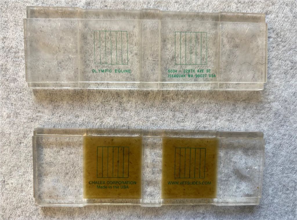
This technique is quantitative and requires the use of specific reusable slides (Figure 1) which can be purchased commercially. Saturated salt solutions are typically the flotation solution of choice for this test. The modified McMaster’s FEC has a sensitivity of 25 or 50 eggs per gram (epg) of feces, which is usually sufficient as lower egg numbers do not typically require clinical detection. Consistency in performing this procedure is crucial.

Credit: Brittany Diehl, DVM, MS, UF College of Veterinary Medicine
List of Materials Needed to Perform McMaster’s FEC
- Flotation solution: As prepared from above.
- Digital scale: Capable of weighing in 0.1-gram increments.
- Plastic zip-top sandwich bags: For holding fecal samples.
- Markers: To label samples.
- Disposable cups: For mixing fecal samples with flotation solution.
- Tea strainer: For straining the fecal mixture.
- Tongue depressors: For mixing and crushing fecal samples.
- 30 cc syringe: For measuring flotation solution.
- 3 cc syringe: For measuring fecal suspension.
- Disposable exam gloves: For handling fecal samples.
- Obstetrical lubricant: For collecting rectal samples.
- Eye dropper: For transferring suspension to the McMaster slide.
- McMaster egg counting slide: For counting parasite eggs.
- Microscope: Capable of 100x magnification with a 10x wide field lens and an internal light source.
- Refrigerator or cooler: To keep samples cool.
Modified McMaster’s Procedure for Ruminants
- Weigh and mix: Weigh 4 grams of feces and mix with 56 mL of flotation solution.
- Strain: Strain the mixture to remove large debris.
- Fill the slide: Avoid producing bubbles. Carefully fill each chamber of the McMaster slide with the strained solution. Each chamber holds about .15 mL of suspension.
- Perform microscopic evaluation: Allow the slide to sit for 5 minutes, then examine it under a microscope. The slide must be evaluated no more than 60 minutes after filling.
- Count and calculate: Count the eggs within the grid lines of both chambers and calculate the eggs per gram (epg) by multiplying the total number of eggs by 50.
- Rinse: When finished, rinse the McMaster’s slide with warm tap water (do not use soap or other cleaning solutions).
This method is for a sensitivity of 50 epg. If a sensitivity of 25 epg is required, 4 grams of feces in 26 mL of flotation solution would be needed instead. The total number of eggs would then be multiplied by 25. This method may be preferred in young ruminants.
Microscopic Evaluation of the Slide
Identification of Eggs and Larvae
Examine the slide under a microscope to identify and count the parasite eggs and larvae present. Use a reference guide to accurately identify different species based on egg morphology.
Each type of parasite should be counted separately. When conducting a pre/posttreatment paired evaluation, it is important to understand that less than 90% reduction and less than 60% reduction in fecal egg counts suggest mild and severe resistance, respectively.
Limitations of Fecal Egg Counts
While FECs are a valuable tool for managing parasite loads in livestock, they have several limitations:
- Detection sensitivity: FECs have a lower sensitivity limit, often failing to detect low levels of parasitic infection that might still be clinically significant.
- Egg shedding variability: Parasite egg shedding can vary significantly depending on the time of day, the individual animal, and the parasite's life cycle stage, leading to inconsistent results.
- Species identification: FECs do not differentiate between species of parasites, especially within the strongyle family, which can complicate treatment decisions.
- Snapshot in time: The results provide a single point-in-time snapshot and may not reflect the overall parasite burden due to daily variations in egg shedding.
- External factors: Factors such as animal stress, nutrition, and immune status can affect FEC results, potentially leading to inaccuracies.
Conclusions
Regular FECs are an essential tool for effective parasite management in grazing livestock. By monitoring parasite burdens and the efficacy of dewormers, producers can make informed decisions to maintain the health and productivity of their animals. It is crucial to integrate FAMACHA® scoring and other visual assessment techniques, such as the Five Point Check©, to supplement FEC data for a comprehensive approach to parasite control. Producers should consult with a veterinarian to interpret FEC results and make informed treatment decisions, as a valid VCPR is necessary for legal and effective parasite management. One size does not fit all regarding parasite management.
References
Fernandez, D. 2012. "Fecal Egg Counting for Sheep and Goat Producers." University of Arkansas at Pine Bluff.
Hale, M. 2015. “Managing Internal Parasites in Sheep and Goats.” ATTRA, National Center for Appropriate Technology. https://attra.ncat.org/publication/managing-internal-parasites-in-sheep-and-goats/
Liotta, J. n.d. “Fecal Egg Counting Procedure.” Cornell Sheep and Goat Symposium, Cornell University. http://goatdocs.ansci.cornell.edu/CSGSymposium/BasicQuantitativeFecalExaminationMethod.pdf
O’Brien, D., K. Matthews, N. Whitley, and S. Schoenian. 2019. "Using Fecal Egg Counts on Your Farm." Virginia Cooperative Extension.
Scott, D. 2018. "How Fecal Egg Counts Can Help You Fight Parasites." ATTRA, National Center for Appropriate Technology. https://attra.ncat.org/publication/how-fecal-egg-counts-can-help-you-fight-parasites/
Storey, B. 2022. "Fecal Egg Counts: Uses and Limitations." University of Georgia College of Veterinary Medicine.
Zajac, A. Z., and G. A. Conboy. 2012. Veterinary Clinical Parasitology. 8th edition. 8–11.