Several different fungi and one bacterium cause leaf spot diseases of strawberry. Symptoms caused by these pathogens are often similar, leading to confusion and misdiagnosis of the disease. This publication focuses on helping growers, crop consultants, and Extension agents with the diagnosis and identification of the most common strawberry leaf spot diseases found in Florida. Generally, most of these leaf spots have not been of significant concern in the strawberry industry and are considered of minor importance. Losses associated with most of these diseases are rarely observed. Therefore, except for powdery mildew and angular leaf spot, no specific control measures and recommendations have been developed for these diseases. Research to develop specific recommendations for Pestalotia leaf spot is currently underway. Table 1 and Figure 1 below summarize the common strawberry leaf spot diseases covered in this publication.
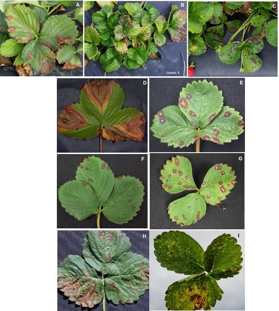
Credit: A, C–I: UF/IFAS GCREC; B: F. Louws
Pestalotia Leaf Spot and Fruit Rot
Pestalotia leaf spot and fruit rot are caused by species of Neopestalotiopsis. Symptoms are characterized by light-to-dark-brown spots of varying sizes, irregularly distributed on infected leaves (Figure 2A). Young spots are often circular but become irregular as they enlarge, especially near the margins of the leaf. In advanced stages, the spots increase in size and may merge. Pestalotia leaf spots can be confused with early stages of leaf blotch (caused by Gnomonia comari), which is also covered in this publication, but are distinguished by black spore masses that form in the tan centers of older spots, especially under humid conditions (Figure 2B).
In recent outbreaks in Florida, severe spotting often produced blight-like browning of older leaves, eventually killing them. The disease can also decline plant vigor, resulting in stunted growth and development of smaller and weaker leaves. In severe cases, the pathogen infects roots and crowns, leading to the eventual death of the plant.
Fruit symptoms can be confused with anthracnose fruit rot (AFR). Like AFR, fruit rot begins as dry, light-tan, slightly sunken, irregularly shaped lesions 2–4 mm in diameter. The lesions expand and may take over the entire fruit. Unlike AFR, however, large lesions are eventually covered by dark fruiting bodies (acervuli), which produce spores in shiny black droplets of liquid (Figure 2C). These symptoms take time to develop in the field but may be hastened by incubating diseased fruit (or leaves) in moist plastic bags for 24 to 48 hours to induce sporulation.
The fungus produces spores on the surface of infected tissues, which can be spread by water and farm operations. Disease development is favored by consecutive or extended rain events and temperatures above 50°F (10°C), with optimum temperatures between 77°F and 86°F (25°C and 30°C). When these conditions are prolonged, epidemics develop, which are very difficult to control.
For more information and specific recommendations, visit https://edis.ifas.ufl.edu/pp357.

Credit: UF/IFAS GCREC
Common Leaf Spot
Common leaf spot, caused by Mycosphaerella fragariae, is also known as Mycosphaerella leaf spot, Ramularia leaf spot, "rust," bird's-eye spot, "gray spotness," or white spot. Symptoms can be confused with early stages of leaf blight (caused by Phomopsis obscurans) and leaf blotch (caused by Gnomonia comari), although the lesions caused by this pathogen do not enlarge as aggressively. It can also be confused with leaf scorch (caused by Diplocarpon earliana), which is covered in the next section.
Symptoms are characterized by leaves with small and circular leaf spots with light to tan centers and purplish borders (Figure 3). Lesions usually start on the upper leaf surface as small, deep purple, round to irregularly shaped necrotic spots, which can grow to 1–2 mm in diameter. On young leaves, spots remain light brown, whereas on older leaves the center changes from tan or brown to gray or white, and the necrotic center is surrounded by reddish-purple to rusty-brown borders. Lesions occur less frequently on the underside of the leaves, and the color is not as vivid. Intense spotting may lead to death of the infected leaves.
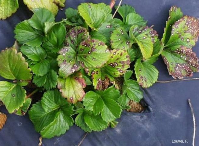
Credit: F. Louws
Symptoms differ based on the strawberry cultivar, strain of the fungus, and temperature range. Uniformly rusty-brown spots without purplish borders or light necrotic centers may be produced on young leaves when warm and humid conditions are present.
M. fragariae also causes spots on fruit, calyxes, petioles, and runners. Under high humidity, superficial black spots (¼ inch across) can be formed on fruit around groups of seeds. Fruit spots and black seeds are usually limited to one or two per fruit.
In Florida, the asexual stage (Ramularia tulasnei) is more common and produces conidia that can be dispersed by splashing water from rains or overhead irrigation. The pathogen is favored by high humidity and temperatures between 50°F and 80°F (10°C and 27°C).
Leaf Scorch
Leaf scorch is caused by the fungus Diplocarpon earliana. Symptoms can be mistaken for common leaf spot, caused by M. fragariae.
Several small, irregular, and purplish to tan spots or "blotches" (1–5 mm in diameter) develop on the upper surface of leaves (Figure 4A). In contrast to common leaf spot (caused by M. fragariae), the centers of the blotches remain purple or slowly develop small brownish centers. When blotches are numerous and start to merge, tissues between the lesions often turn bright red to brown (Figure 4B). In severe cases, entire leaves curl up at the margins, turn brown, and acquire a burned (scorched) appearance.
D. earliana also infects petioles, flowers, and fruit. Infected sepals and petioles develop irregular purple to brown spots (Figure 4C). Badly spotted sepals may turn brown, resulting in "dead caps," which make fruit unattractive and lower the market grade. In severe cases, infected flowers and fruit may die.
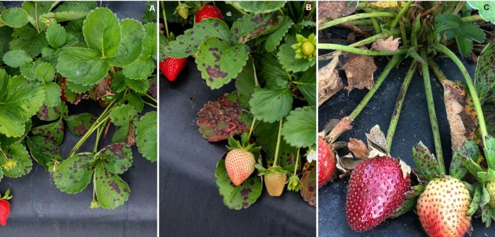
Credit: UF/IFAS GCREC
Leaf scorch is favored by long periods of leaf wetness, frequent rain, and temperatures between 60°F and 77°F (15°C and 25°C). Older and middle-aged leaves are infected more easily than young ones. Disease occurrence is common when fungicides labeled for strawberry production are not applied regularly. Applications of captan and other strawberry fungicides seem to be effective in reducing disease incidence.
Phomopsis Leaf Blight and Soft Rot
Phomopsis leaf blight and soft rot is caused by the fungus Phomopsis obscurans. Symptoms start as one to numerous circular reddish-purple lesions on a leaflet. During the early stages, symptoms are practically indistinguishable from common leaf spot lesions, caused by M. fragariae, due to the development of gray centers in the spots. As the disease progresses, spots enlarge to V-shaped lesions with light-brown centers and darker-brown outer zones. Lesions follow major veins progressing inward (Figure 5A). The fungus produces black fruiting bodies (pycnidia) on the inner zone of the lesion. If pycnidia are not present, leaves may be incubated in a moist chamber for 24 to 48 hours to induce sporulation.
The fungus also infects petioles, runners, calyxes, and fruit. Pink and red fruit are usually affected, and initial symptoms are characterized by round, light-pink, and water-soaked lesions. In more advanced stages, lesions turn light brown at their margins and darker brown and crusty toward the centers, covered with several black pycnidia (Figure 5B).
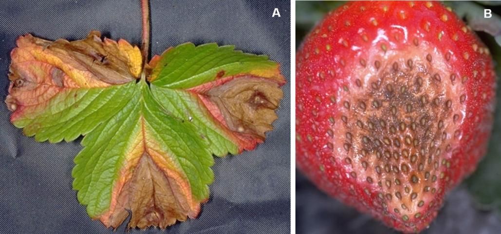
Credit: UF/IFAS GCREC
The pathogen is usually introduced with transplants, and the disease might build up in fruit production fields, but it is rarely a concern among growers. The disease is favored by humid weather, and the pathogen is dispersed by water splash from rains or overhead irrigation.
Leaf Blotch and Stem-End Rot
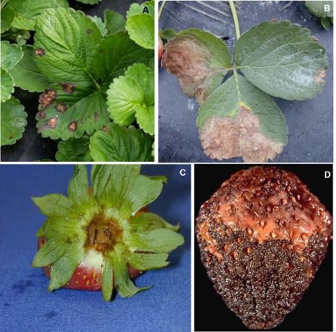
Leaf blotch, caused by Gnomonia comari, may be confused with several other leaf spot diseases, especially when lesions are young. Young spots are brownish and roughly circular with purple borders (Figure 6A). The spots expand aggressively over time, often forming large light brown necrotic areas. Such areas may develop faint zonate patterns and often extend to the margins of the leaf (Figure 6B). Fruiting bodies (pycnidia) form on old spots as upraised tan to brown structures whose color resembles surrounding tissues. As a result, they may not be readily visible. If pycnidia are not visible with a hand lens, leaves may be incubated in a moist chamber for 24 to 48 hours to induce sporulation. Old outer leaves often die from a combination of leaf spotting and infection of the lower petioles.
The pathogen also infects flowers, calyxes, and fruit, causing stem-end rot (Figure 6C). Irregular brown areas form on green fruit, causing them to ripen prematurely or dry up, inhibiting further development. Symptoms on older fruit are characterized by irregular brown lesions, generally expanding from under the cap. Soft rot symptoms are produced on ripe fruit, which can be covered with dark fruiting bodies (Figure 6D).
Credit: UF/IFAS GCREC
Leaf blotch and stem-end rot occur sporadically and are often associated with the nursery source of the transplants. Disease development is favored by overhead irrigation and frequent rain. Currently grown cultivars such as Florida127 (Sensation™) and Florida Brilliance seem particularly prone to the leaf spot phase of this disease. G. comeri is considered a weak pathogen and usually infects plants through wounds and stomata on the leaves.
Cercospora Leaf Spot
Cercospora leaf spot is caused by the fungus Cercospora fragariae. In the early stages, leaf spots are small, circular, and uniformly purplish/reddish with very light-colored (almost white) centers, apparent only on the upper surface (Figure 7A). The spots resemble common leaf spot (caused by M. fragariae) but are smaller with lighter centers and more irregular shapes.
In advanced stages, the necrotic centers become brittle and may fall out, resulting in "shot hole" symptoms (Figure 7B). The spots remain relatively small and irregular with a more defined dark-purple outer zone, and they occasionally coalesce. On the lower leaf surface, the spots are bluish to tan in color and appear more diffuse. Spore production occurs on the upper surface of the leaf, in the white centers which become dotted with small dark stroma or knots of fungal cells (Figure 7C). Like many fungal pathogens, C. fragariae is spread by water splash. However, it does not infect the fruit and is considered a minor strawberry pathogen.

Credit: UF/IFAS GCREC
Target Spot
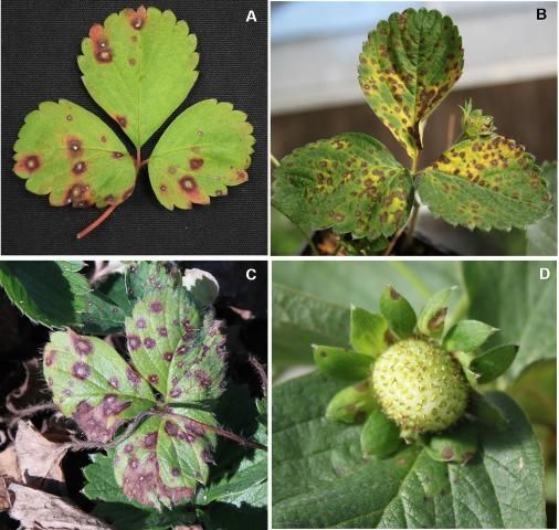
Target spot is caused by the fungus Corynespora cassiicola. Early symptoms are characterized by small (3 to 5 mm across), circular to irregular spots on leaves with dark brown borders and beige centers (Figure 8A). When spotting is intense, leaf lesions can expand to 20 mm, and surrounding leaf tissues may become yellow (Figure 8B). In severe infections, spots can occupy half of the leaf area (Figure 8C). Symptoms can also be observed on petioles and the fruit calyx (Figure 8D).
Credit: UF/IFAS GCREC
This disease has been reported recently on strawberry and seemed associated with nursery transplants that were infected by another host in close-by fields.
Powdery Mildew
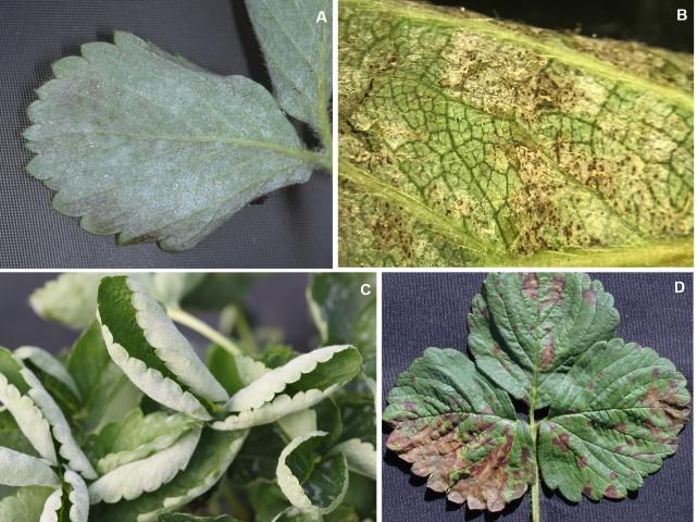
Strawberry powdery mildew is caused by the fungus Podosphaera aphanis. The disease is easily recognized by the appearance of fuzzy white fungal growth on lower leaf surfaces (Figure 9A). Small black fruiting bodies (cleistothecia) may develop on the undersides of heavily infected leaves (Figure 9B). Leaf cupping is common and occurs when the margins of diseased leaves curl upward and inward (Figure 9C). In some cultivars, irregular red or purple/brown patches develop on one or both leaf surfaces (Figure 9D), which can be confused with other leaf spot diseases. Mycelial growth also occurs on flowers and fruit, especially those embedded in the plant canopy, as well as on the upper leaf surface of plants growing in tunnels and greenhouses.
Credit: UF/IFAS GCREC
P. aphanis is carried on infected transplants, and it is not believed to persist over our hot, wet Florida summers. Spore germination and fungal growth are favored by mild temperatures between 60°F and 77°F (15°C and 25°C), high relative humidities (75% to 98%), and rapid vegetative growth, which may explain why epidemics often occur between November and January. High temperatures, strong sunlight, heavy rains, and wet leaf surfaces suppress disease development. Powdery mildew is readily controlled by various fungicides applied at 2-week intervals, but applications must be started early in the season (late November), before fungal growth is extensive and leaf curling becomes obvious.
For more information and specific management recommendations, visit https://edis.ifas.ufl.edu/pp129.
Angular Leaf Spot
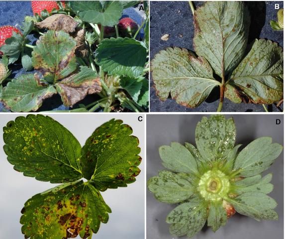
Angular leaf spot (ALS) is a bacterial disease caused by Xanthomonas fragariae. Leaves are the primary target tissue, although leaflike sepals on the fruit caps, known as fruit calyx, may also be infected. Mass spotting is common during outbreaks in Florida, resulting in yellowing, blighting, and premature death of older leaves (Figure 10A). Spots are small in size (3–5 mm across) and confined by leaf veins, giving them a blocky or angular shape. Young spots look water-soaked (Figure 10B) but appear translucent when held up to the light (Figure 10C). This "windowpane" effect is diagnostic for ALS. Individual spots may also coalesce and run together along the larger veins of a leaf. Older spots turn brown to black with age. When environmental conditions are favorable, sepals are also infected. In severe cases, mass spotting of the sepals results in "brown cap" symptoms, rendering the fruit unmarketable (Figure 10D).
Credit: UF/IFAS GCREC
ALS outbreaks are common in some seasons but not others. Epidemics usually occur when rains or overhead irrigation for frost protection are frequent. During wet, humid weather, bacteria ooze from the spots on the lower leaf surface and are spread by splashing water, farm equipment, and harvesters. ALS is not controlled by fungicides but is suppressed by copper-containing products, as well as the plant defense promoter Actigard. Unfortunately, these products also tend to suppress yield when applied too frequently or at high rates.
For more information and specific management recommendations, visit https://edis.ifas.ufl.edu/pp120.
References
Ellis, M. A. 2016. Strawberry Leaf Diseases. Ohioline. PLPATH-FRU-35. The Ohio State University Extension. Plant Pathology. https://ohioline.osu.edu/factsheet/plpath-fru-35
Louws, F., G. Ridge, and B. Cline. 2019a. Gnomonia Leaf Blotch and Stem-End Rot of Strawberry. NC State Extension Publications. https://content.ces.ncsu.edu/gnomonia-comari-leaf-blotch-of-strawberry
Louws, F., G. Ridge, and B. Cline. 2019b. Phomopsis Leaf Blight of Strawberry. NC State Extension Publications. https://content.ces.ncsu.edu/phomopsis-leaf-blight-of-strawberry#
Maas, J. L. 1998. Compendium of Strawberry Diseases, 2nd edition. St. Paul: APS Press.
Onofre, R. B., C. S. Rebello, J. C. Mertely, and N. A. Peres. 2019. "First Report of Target Spot Caused by Corynespora cassiicola on Strawberry in North America." Plant Dis. 103:1412.