This EDIS publication provides information to seafood consumers on the differences between white and red fish fillets' composition and nutritional value, as well as the effect of heat on color change while cooking. Note that the term “muscle” and “fillet” are used interchangeably in this publication, however there are some differences in common usage.
Introduction
Fish, as one of the primary sources of protein and other nutrients, comes in a diverse array of shapes, sizes, and muscular systems (Johnston 1983). According to the Food and Agriculture Organization of the United Nations (FAO 2002), fish provides almost 16% of the animal protein consumed by the world’s population. This popularity is not only due to the health benefits that fish offers but also its unique sensory characteristics such as flavor, color, odor, and texture. Variations in chemical composition may lead to changes in these attributes that determine the acceptability of fish fillets as food (Venugopal and Shahidi 1996). This EDIS publication aims to provide information on the differences between red muscles and white muscles in fish, as well as fish muscle structure and nutritional composition. Furthermore, the effect of heat on the muscle and its relation to some of the sensory characteristics are briefly discussed.
Structure of Fish Muscle
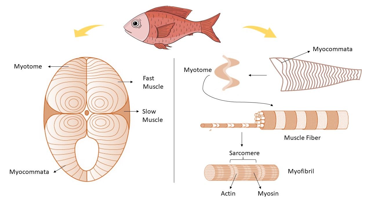
In fish, the fillet consists of two bundles of large lateral muscles that run on both sides of the backbone. "Myocommata" which is a thin connective tissue membrane, divides fish muscle into muscle blocks known as “myotomes” (Figure 1). Each myotome is composed of muscle fibers generally less than 20 mm long and 0.02–1 mm in diameter (Venugopal and Shahidi 1996). Each muscle fiber contains 1000–2000 “myofibrils,” with diameters ranging between 1–2 µm. The myofibril consists of small subunits named “sarcomeres” (Belitz, Grosch, and Schieberle 2009). Sarcomeres are composed of the main contractile proteins, namely, actin (thin filament) and myosin (thick filament). The intramuscular connective tissues mostly consist of sheets of collagen. The collagen content directly affects the textural properties of the fish muscle, such as firmness (Suárez et al. 2005). This explains why fish fillet is more digestible compared to mammalian muscle, mainly because there is less intramuscular connective tissue in fish (Abraha et al. 2018; Alam 2007). Also, the length of the muscle fibers and the myocommata’s thickness increases as fish get older (Listrat et al. 2016), so it is expected that older fish have firmer muscles.
What differentiates fish from terrestrial animals is the anatomical separation of the three main types of muscle: white muscle (also known as fast muscle), a superficial red muscle (slow muscle), and a pink muscle (intermediate) (Alam 2007) which will be further discussed in the next section.

Credit: Rose Omidvar and Razieh Farzad, UF/IFAS
Differences Between Red and White Fillet
Fish can be categorized in many ways. The body form (e.g., round, flat), their habitat (e.g., sea, freshwater), and the type of muscle they have (e.g., white, red) are some examples (Belitz et al. 1970). In this section, the latter will be discussed further.
Most fish have a combination of different types of muscles, each with different physiological roles. The difference in color depends on the amount of myoglobin (an oxygen-carrying protein in muscle) as well as the type of food the fish consumes (Okwuosa, Amadi-Ibiam, and Omovwohwovie 2021).
Red Muscle
The red muscle usually lies directly under the skin along the side of the body and, in certain active species, also in a band near the spine. The red muscle is similar to the heart muscle in that it is rich in mitochondria, is well supplied with capillaries, has a higher content of myoglobin, and hence has greater oxygen availability (Belitz, Grosch, and Schieberle 2009). This makes red muscles suitable for slow and continuous swimming (Listrat et al. 2016). Nearly 48% of the body weight of persistent-swimming fish species, like herring or mackerel, consists of red muscle (Belitz, Grosch, and Schieberle 2009). The red muscle cells primarily rely on aerobic metabolism (using oxygen) to provide energy for consistent muscle work and are richer in lipids, nucleic acids, myoglobin, and vitamin B compared to white muscle cells (Alam 2007).
White Muscle
Depending on the activity level of the fish and the speed required for swimming, the proportion of red to white muscles varies. With increasing speed, the involvement of the white muscle increases as well. Compared to red muscle, white muscle has thicker fibers and possesses fewer capillaries leading to less blood flow, hence reduced oxygen availability. These muscles contract rapidly and less sustainably, having an anaerobic metabolism (not using oxygen). This attribute makes them suitable for sudden, quick movements needed for escaping from a predator or for catching prey before they exhaust their supply of glycogen and need to rest. The mitochondria density in white muscle cells is approximately five times less than in red muscle cells (Moyes and Hood 2003). Most of the fish found in freshwater that are a slow-moving or bottom-feeding type have predominantly white muscles (Alam 2007).
Pink Muscle
Also known as intermediate muscle, pink muscle has an intermediate functionality of white and red muscle, which makes it suitable for continued swimming efforts lasting a few tens of minutes at a moderately high speed. However, the pink color found in salmon and sea trout does not stem from myoglobin content. Since those fish feed on crustaceans, they develop a pink color due to the red carotenoid called astaxanthin. Fish are incapable of synthesizing astaxanthin, so the degree of pink color in the muscle depends on the consumption of pigmented diet (Alam 2007).
Nutritional Composition of Fish
Fish is an excellent source of macronutrients (e.g., protein, carbohydrates, and lipids) and micronutrients (e.g., vitamins and minerals). It is well known for having highly digestible protein and is considered a great source of omega‐3 fatty acids (Ahmed et al. 2022).
Omega-3 fatty acid is an essential fatty acid, meaning the body cannot make it from scratch and needs to obtain it through diet. Consumption of omega-3 fatty acids is of great importance because not only do they provide energy for the body but they are also an integral part of cell membranes, affecting the function of cell receptors there (SanGiovanni and Chew 2005). Other health benefits associated with omega-3 consumption are the prevention and treatment of cardiovascular, inflammatory, and neurological diseases such as Alzheimer’s disease (Abraha et al 2018).
Fish contains the approximate chemical composition of 75% water, 20% protein, 1–10% lipid, 1–2% minerals, and 0.1–1% carbohydrate (Listrat et al. 2016). This nutritional composition varies according to species, size, age, feeding habits, water temperature, etc. (Ahmed et al. 2022). Table 1 shows the average composition of various fish as a percentage of edible portion and the gram of omega-3 content per 100 grams of the fillet reported (Belitz, Grosch, and Schieberle 2009).
Table 1. Average composition of various fish as % of edible portion and omega-3 content as (g/100 g of fillet).
It is worth noting that the chemical composition of white and dark muscle within the same species can vary as well. For example, Table 2 reports that the red and white muscles of pink salmon and albacore tuna exhibit a difference in the proximate composition. As previously mentioned, these muscles play a different physiological role in fish (Ahmed et al. 2022).
Table 2. Composition of light and dark pink salmon and tuna albacore.
Why do the color and the texture of the fish fillet change after cooking?
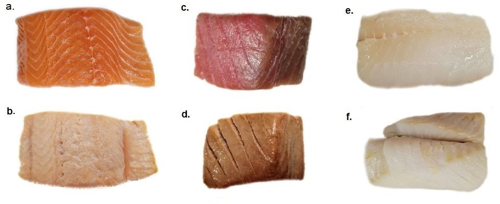
Heating is a fish processing method that enhances the taste and flavor as well as prolongs the shelf life of fish. Heat can be applied in different ways, such as boiling, baking, roasting, frying, and grilling (Abraha et al. 2018). Upon heating, the muscle becomes opaque and firm (Hultin 1984). In addition, the collage network gelatinizes and results in the disintegration of muscle tissue into a flake-like texture (Belitz, Grosch, and Schieberle 2009). The conformational changes in proteins that happen when they are heated are referred to as denaturation. Thermal denaturation of muscle involves the unfolding of protein (especially contractile protein) molecules, which occurs due to the breakage of intermolecular hydrogen bonds, thus reducing the water binding capacity and shrinkage of the muscle (Yu et al. 2014). Two of the main factors during heat processing are time and temperature, which can affect protein structure and sensory qualities. The increase in time and temperature results in higher degrees of protein denaturation (Abraha et al. 2018). Heating not only denatures the muscle proteins but also the muscle pigment and predisposes it to oxidation. The oxidization of the iron in the heme group produces metmyoglobin which, upon heating, denatures and creates brown color (Hultin 1984). Figure 2 below shows the color changes of three different fish fillets after cooking.

Credit: Rose Omidvar and Razieh Farzad, UF/IFAS
References
Abraha, B., H. Admassu, A. Mahmud, N. Tsighe, X. W. Shui, and Y. Fang. 2018. “Effect of Processing Methods on Nutritional and Physico-chemical Composition of Fish: A Review.” MOJ Food Processing & Technology 6 (4): 376–382. https://doi.org/10.15406/mojfpt.2018.06.00191
Ahmed, I., K. Jan, S. Fatma, and M. A. O. Dawood. 2022. “Muscle Proximate Composition of Various Food Fish Species and Their Nutritional Significance: A Review.” Journal of Animal Physiology and Animal Nutrition 106 (3): 690–719. https://doi.org/10.1111/jpn.13711
Belitz, H. D., W. Grosch, and P. Schieberle. 2009. “Fish, Whales, Crustaceans, Mollusks.” In Food Chemistry. 617–639. Germany: Springer, Berlin, Heidelberg. https://doi.org/10.1007/978-3-540-69934-7_14
Food and Agriculture Organization of the United Nations (FAO). 2002. “3. Global and Regional Food Consumption Patterns and Trends.” In Joint WHO/FAO Consultation on Diet, Nutrition, and the Prevention of Chronic Diseases. Retrieved on March 13, 2023. https://www.fao.org/3/ac911e/ac911e05.htm
Hultin, H. O. 1984. “Postmortem Biochemistry of Meat and Fish.” Journal of Chemical Education: 61 (4): 289. https://doi.org/10.1021/ed061p289
Johnston, I. A. 1983. “On the Design of Fish Myotomal Muscles.” Marine Behaviour and Physiology 9 (2): 83–98. https://doi.org/10.1080/10236248309378586
Listrat, A., B. Lebret, I. Louveau, T. Astruc, M. Bonnet, L. Lefaucheur, B. Picard, and J. Bugeon. 2016. “How Muscle Structure and Composition Influences Meat and Flesh Quality.” The Scientific World Journal 2016: 3182746. https://doi.org/10.1155/2016/3182746
Moyes, C. D., and D. A. Hood. 2003. “Origins and Consequences of Mitochondrial Variation in Vertebrate Muscle.” Annual Review of Physiology 65: 177–201. https://doi.org/10.1146/annurev.physiol.65.092101.142705
Alam, A. N. 2007. “Structure of Fish Muscles and Composition of Fish.” In Participatory Training of Trainers: A New Approach Applied in Fish Processing. 37–48. Bangladesh Fisheries Research Forum. https://www.researchgate.net/publication/342231886
Okwuosa, O. B., C. O. Amadi-Ibiam, and O. E. Omovwohwovie. 2021. “A Review on Fish Growth and Physiological Properties of Fish Muscle Tissue Development.” IRE Journals 5 (6): 20–29. https://www.irejournals.com/formatedpaper/1702990.pdf
SanGiovanni, J. P., and E. Y. Chew. 2005. “The Role of Omega-3 Long-Chain Polyunsaturated Fatty Acids in Health and Disease of the Retina.” Progress in Retinal and Eye Research 24 (1): 87–138. https://doi.org/10.1016/j.preteyeres.2004.06.002
Suárez, M. D., M. Abad, T. Ruiz-Cara, J. D. Estrada, and M. García-Gallego. 2005. “Changes in Muscle Collagen Content During Post Mortem Storage of Farmed Sea Bream (Sparus aurata): Influence on Textural Properties.” Aquaculture International 13 (4): 315–325. https://doi.org/10.1007/s10499-004-3405-6
Venugopal, V., and F. Shahidi. 1996. “Structure and Composition of Fish Muscle.” Food Reviews International 12 (2): 175–197. https://doi.org/10.1080/87559129609541074
Yu, X., Y. Llave, M. Fukuoka, and N. Sakai. 2014. “Estimation of Color Changes in Fish Surface at the Beginning of Grilling Based on the Degree of Protein Denaturation.” Journal of Food Engineering 129: 12–20. https://doi.org/10.1016/j.jfoodeng.2013.12.030