Introduction
Industrial hemp is Cannabis sativa that contains < 0.3% THC and can be legally grown with permits in Florida. The plant may be grown for flower, grain, or fiber production. Other than the lower THC levels, hemp is the same plant botanically as marijuana. Because this plant has long been on the Schedule 1 controlled substance list in the U.S. (United States), knowledge of pests and diseases is limited, and labeled control measures are almost non-existent. This publication provides information and general management recommendations for hemp diseases found in Florida. Management options are currently limited as there is a lack of resistant varieties and other management strategies for industrial hemp production.
For more information on hemp diseases including updated pesticide product lists, refer to the link https://plantpath.ifas.ufl.edu/extension/hemp-diseases/
Disease Identification
For help with identifying hemp diseases, consider submitting samples to the UF/IFAS Plant Diagnostic Center for pathogen identification and confirmation of disease.
More information on the different Plant Disease Clinics and their submitting processes as well as sample collection and shipping instructions can be found here: https://plantpath.ifas.ufl.edu/extension/diagnostic-labs/
Hemp Permits may be required for submitting plant samples to the disease clinic. Please check with the clinic before sending your samples.
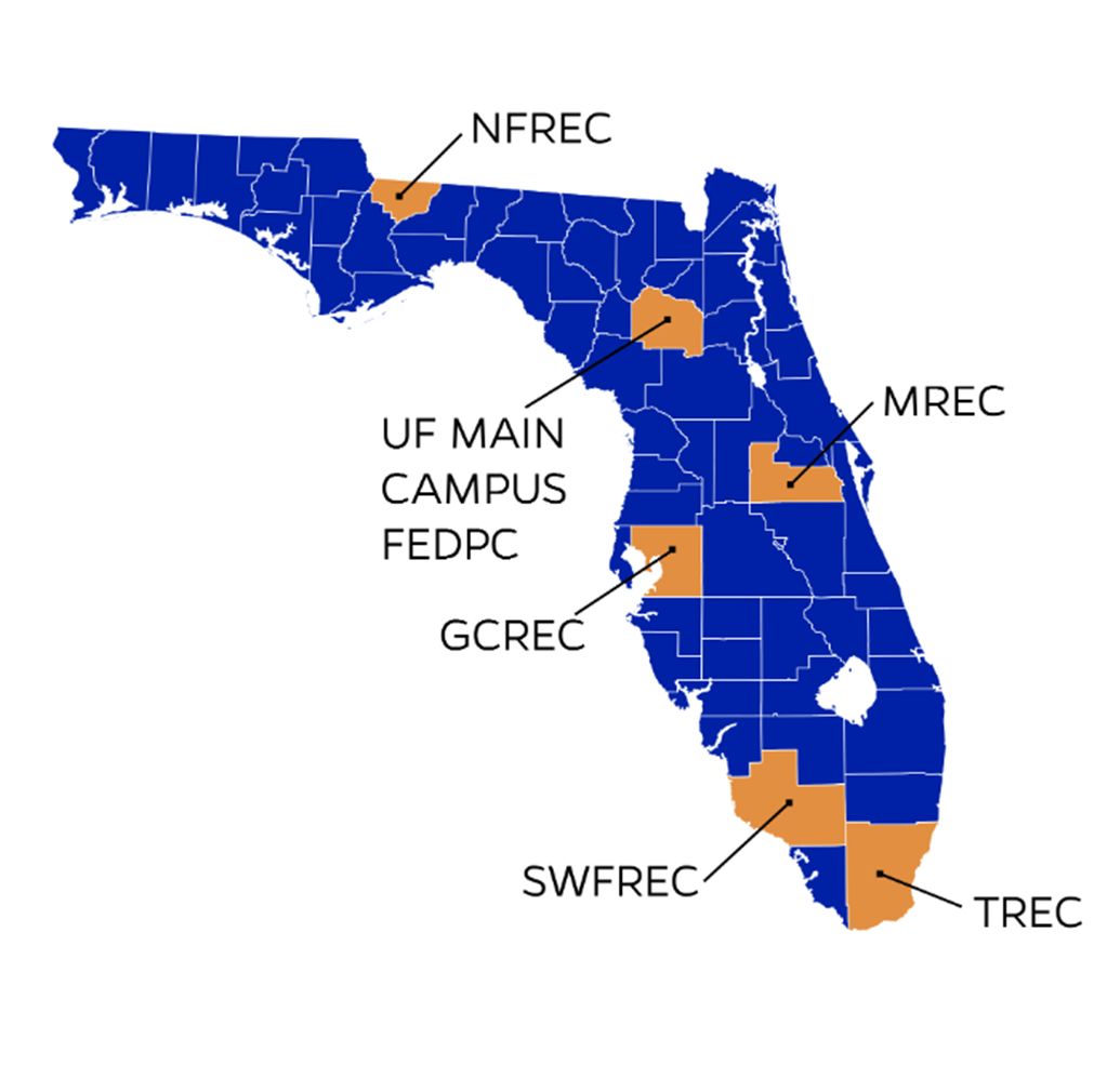
Credit: UF/IFAS
Plant Disease Clinic – Main Campus
2570 Hull Rd, Gainesville, FL 32603
GCREC Plant Disease Clinic
14625 CR 672, Wimauma, FL 33598
NFREC Plant Disease Clinic
155 Research Road, Quincy, FL 32351
TREC Plant Disease Clinic
18905 SW 280th St., Homestead, FL 33031
Leaf Spot Diseases
Bipolaris Leaf Spot – Bipolaris sp.
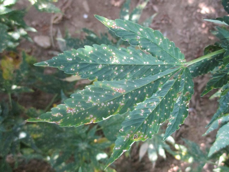
Credit: Thomas Isakeit, Texas A&M University
Signs & Symptoms
Small, circular, light green spots develop on the leaves. As the disease progresses, the spots expand to around 1- to 2-mm in diameter turning brown with a dark border. Lesions may also remain light tan and lack a border. Several lesions may coalesce to form a larger, irregularly shaped lesion. The fungus will sporulate and create dark gray or black “fuzz” on both the top and bottom leaf surfaces. This can be viewed with a hand lens. Lesions can expand to blight an entire leaf, and necrotic leaves will remain attached to the stem. In severe cases, yield loss can reach 100% (Szarka et al. 2020).
Disease Cycle
The fungus survives in infected plant residue, and rain splashes spores onto healthy plants in the spring. Spores germinate and can infect the leaves and stems of hemp plants. This pathogen can produce multiple infections per growing season (polycyclic). Several weed species have been found to host this fungus which helps keep it in and around fields.
Cercospora Leaf Spot – Cercospora cf. flagellaris
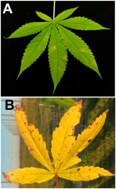
Credit: Marcus Marin, Jacqueline Coburn, Johan Desaeger, and Natalia Peres; UF/IFAS GCREC
Signs & Symptoms
Symptoms start on older foliage and spread to the rest of the canopy. Cercospora can be confused with Curvularia and/or Septoria leaf spots. Leaf spots start as yellow flecks which expand into light tan or white lesions surrounded by yellow halos. Sporulating structures (conidiophores) at the center of the spots can be visible to the naked eye. Under severe disease conditions, the leaves turn yellow and fall off. Symptoms may progress rapidly and reach up to 60% of the foliage depending on the variety and severity of the disease (Marin, Coburn, et al. 2020).
Disease Cycle
Spores and other infectious fungal parts survive in infected plant residue. Spores are rain-splashed and wind-blown onto the lower parts of plants. Warm and wet conditions in the spring encourage spore germination. Infection is encouraged by leaf wetness.
Curvularia Leaf Spot – Curvularia pseudobrachyspora
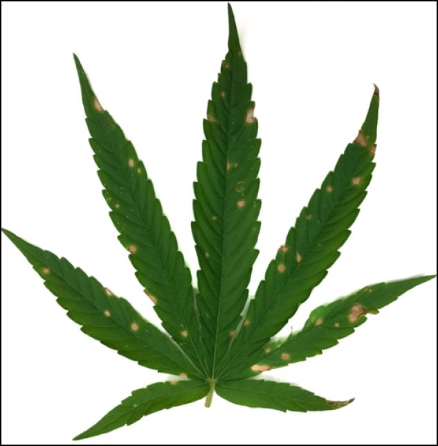
Credit: Marcus Marin, Jacqueline Coburn, Johan Desaeger, and Natalia Peres; UF/IFAS GCREC
Signs & Symptoms
Curvularia leaf spots have a typical appearance of a leaf spot disease and appear on young and old hemp leaves. Symptoms start as yellow spots that turn tan to brown surrounded by a yellow halo as the disease progresses. Symptoms of Curvularia leaf spot can be confused with Cercospora or Septoria. Leaf spot symptoms have been observed to reach 70% incidence depending on the variety (Marin, Wang, et al. 2020).
Disease Cycle
Although more studies are needed to fully understand the epidemiology of this disease, it can be assumed that its life cycle reflects that of most residue-borne, foliar, and fungal diseases. Spores and other infectious fungal parts survive in infected plant residue. Spores are rain-splashed and wind-blown onto the lower parts of plants. Warm and wet conditions in the spring encourage spore germination. Infection is encouraged by leaf wetness.
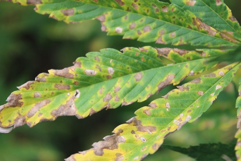
Septoria Leaf Spot – Septoria cannabis
Credit: Nicole Gauthier, University of Kentucky
Signs & Symptoms
Spots start at the older/lower foliage and spread to the rest of the canopy. Symptoms start as irregular light brown spots that turn brown surrounded by a bright yellow halo. Spots can be ¼ inch. As disease severity increases, spots expand and coalesce into brown and yellow lesions that can cause leaf drop. Symptoms can be confused with those caused by Curvularia and/or Cercospora leaf spots. The fungal sporulating structures (pycnidia) can be seen as black pinhead-sized dots in the middle of the lesion. Rapid disease development early in the growing season can reduce overall plant vigor due to foliage loss, by up to 90% (Gauthier et al. 2021).
Disease Cycle
Spores and other infectious fungal parts survive in infected plant residue. Spores are rain-splashed and wind-blown onto the lower parts of plants. Warm and wet conditions in the spring encourage spore germination. Infection is encouraged by leaf wetness, often developing first in the inner canopy and lower leaves where leaf wetness is higher.
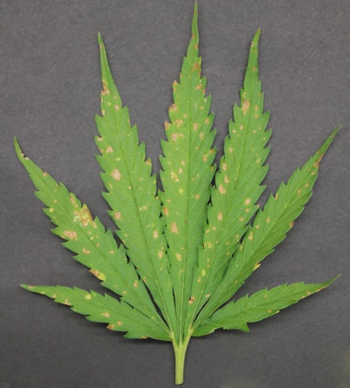
Downy Mildew – Pseudoperonospora cannabina
Credit: Katherine E. M. Hendricks, UF/IFAS SFREC
Signs & Symptoms
Irregular brown to blackish spots can be observed on the lower surface of the leaves with corresponding yellow necrotic spots on the upper leaf surface. With favorable environmental conditions, a purplish to gray mycelial growth may be observed on the lower leaf sides (Giles et al. 2023).
Disease Cycle
Spores and other infectious fungal parts survive in infected plant residue. Spores are rain-splashed and wind-blown onto the lower parts of plants. Cool (< 65°F) and wet conditions in the spring encourage spore production. Infection is encouraged by leaf wetness.
Leaf and Bud Blights
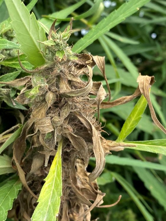
Gray Mold – Botrytis cinerea
Credit: Christian Christensen, UF/IFAS Extension
Signs & Symptoms
Gray mold is one of the common diseases of hemp in Florida due to high humidity. Flower buds are the first to become blighted. Other symptoms such as girdling and breaking of the stem as well as die-back may be observed with increased disease severity. Gray or gray-whitish mycelia and conidia (infectious spores) occur on buds, petioles and stems. Other symptoms such as girdling and breaking of the stem and die-back may be observed with increased disease severity (Garfinkle 2020).
Disease Cycle
The fungus Botrytis cinerea produces sclerotia (hardened survival structures) when host plant resources are depleted. Sclerotia survive in the soil and become infectious in the spring when warm, wet conditions return. Sclerotia germinate to form hyphal mats which give rise to infectious spores. Spores are rain-splashed and wind-blown onto the lower parts of plants. Infection occurs on leaves and buds through wounds and natural openings in the flowers or foliage.
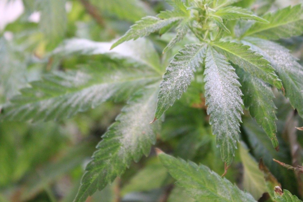
Powdery Mildew – Golovinomyces sp.
Credit: Nicole Gauthier, University of Kentucky
Signs & Symptoms
Fungal growth (mycelia and spores) appears as whitish powdery spots or patches on both sides of the leaves. This distinctive powdery fungal growth can develop quickly (within a week) and can spread across the foliage (leaves, petioles, stems, flower bracts, and buds). Leaf necrosis and defoliation can occur as disease severity increases.
Disease Cycle
Spores and other infectious fungal parts survive in infected plant residue. Spores are rain-splashed and wind-blown onto the lower parts of plants. Warm and wet conditions in the spring encourage spore germination. Unlike most other foliar fungal diseases, powdery mildew infections do not require leaf wetness to occur. Infection is favored by high humidity.
Rust – Uredo kriegeriana
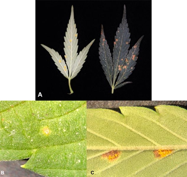
Credit: Zachariah Hansen, University of Tennessee Extension
Signs & Symptoms
Hemp rust infection results in orange, brown, or rust-colored lesions on the top and bottom side of the leaf. Initially, lesions start out light orange and turn dark orange as the disease progresses. Eventually, a mass of orange spores will erupt from the pustules on the bottom side of the leaf.
Disease Cycle
Rust fungi typically require two plant hosts to complete their life cycle. However, the alternate host for hemp rust is not known. Rust spores are wind-blown and deposited on leaf surfaces where they actively penetrate the leaf. Little is known about the life cycle and epidemiology of this disease.
Fungal Diseases of Seedlings, Roots, and Stems
Fusarium Wilt and Crown Rot – Fusarium spp.
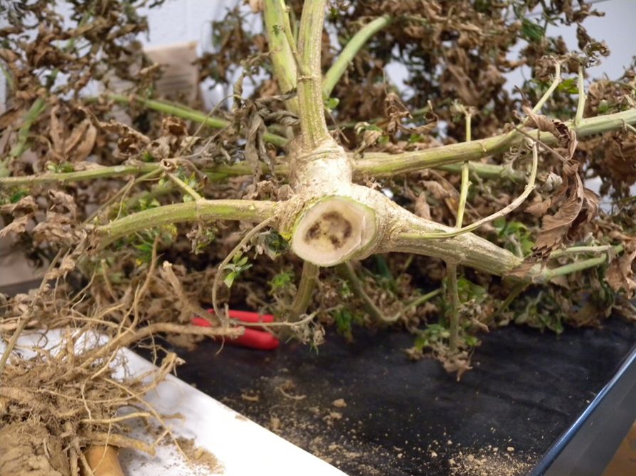
Credit: Brenda Kennedy, University of Kentucky
Signs & Symptoms
Multiple Fusarium species can infect hemp. Fusarium oxyxporum f. sp. vasinfectum has been identified in Florida based on sequencing by F. B. Iriarte at UF-NFREC. Plants will appear chlorotic, stunted, and wilted, especially during the hottest hours of the day. Brown lesions may form on the outside of stems toward the crown. A cross-section of the stem at the crown may reveal brown discoloration of the vascular tissue in severely infected plants. If the infection is severe, roots will appear sunken and brown, or even black. Overall root mass will be less compared to healthy plants.
Disease Cycle
Fusarium is a soilborne fungus and can survive outside of infected plant residue in the soil for long periods. Fusarium can enter a field through machinery, seeds, transplants, and any infested soil. Infections typically occur through the roots. The fungus can move upward through the plant and into the crown and stems, eventually causing complete plant collapse.
Pythium Root, Crown, and Stem Rot – Pythium spp.
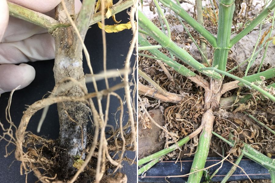
Credit: Shouhua Wang, Nevada Dept. of Agriculture
Signs & Symptoms
Multiple Pythium species are reported to cause root, crown, and stem rot. Symptoms are similar between species. Pythium aphanidermatum and P. myriotylum have been identified in Florida based on sequencing at UF-NFREC 2020. Rot symptoms start as necrosis of smaller feeder roots that extend to larger roots and eventually, the crown, which turns brown to black. Roots will appear soft and slimy. Pythium only infects the root cortex and not the inner root stele, so the outer part of the root may be easily stripped off, leaving the white inner stele. Foliar stress symptoms such as leaf scorching or stunting may be noticed as disease severity increases; this is due to a compromised root and/or vascular system.
Disease Cycle
Pythium is a soilborne fungus-relative and can survive outside of infected plant residue in the soil for extended periods. Pythium is classified as a water mold, and infection by these pathogens is often favored by wet soil conditions and cool temperatures. They can produce motile spores which can “swim” in saturated soil toward plant roots. Initial infection occurs through the root cortex and can progress upward to the crown and stem.
Hemp Canker – Sclerotinia sclerotiorum
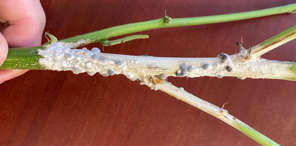
Credit: Christian Christensen, UF/IFAS Extension
Signs & Symptoms
White, cottony mycelial growth appears on the stems. Hard, dark-colored sclerotia start forming on top of the white mycelia as the disease progresses. The sclerotia continue to darken to a blackish color as they mature.
Disease Cycle
The fungus survives by producing dark, hardened masses called sclerotia, that can be found in infected plant debris, in seed lots, and in the soil. Sclerotia can persist in the soil for up to five years. When environmental conditions are right (adequate moisture and temperatures between 41°F – 68°F) apothecia will form which releases infectious spores. Infection occurs mostly on plant parts that are injured and/or dying but can also occur on the stem at the soil line. Like most fungal diseases, infection is favored by wet foliage. This pathogen has a large host range (over 600 plant species) including several agronomic row crops like peanut and soybean.
Southern Blight – Agroathelia rolfsii (formerly Sclerotium rolfsii)
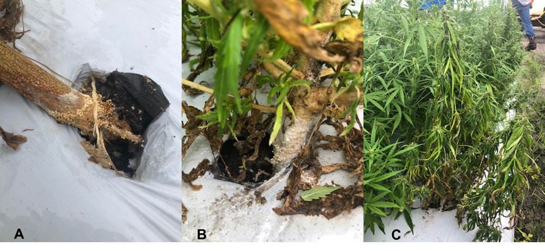
Credit: Evelyne (Prissy) Fletcher, UF/IFAS Extension
Signs & Symptoms
Severe wilting is the primary symptom of southern blight. Leaves of wilted plants may remain green but can also turn yellow. Cottony, whitish mycelia and tan to deep brown sclerotia (mustard seed sized) may be observed on the crown and lower stem of the plant. They may also be observed on dead plant material on the surrounding soil surface. Sometimes during warmer temperatures, the disease will develop as a discolored crown without any other signs or symptoms of the pathogen. Wilted plants may occur adjacent to healthy plants.
Disease Cycle
A. rolfsii is a soil-borne fungus that produces sclerotia (hardened survival structures) when host plant resources are depleted. Sclerotia survive in the soil and become infectious in the spring when warm, wet conditions return. The pathogen also survives as mycelium (fungal strands) on and in dead plant material. Sclerotia germinate to form hyphae which directly infect plant stems, roots, or crowns at the soil line. The optimum temperature range for disease development is 81°F to 95°F (Pfeufer et al. 2018).
Stem Canker – Diaporthe phaseolorum
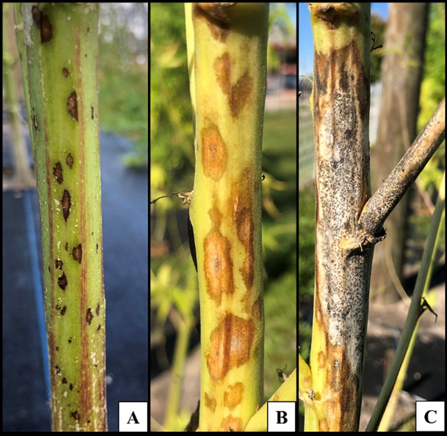
Credit: Marcus Marin, Nan-Yi Wang, Jacqueline Coburn, Johan Desaeger, and Natalia Peres; UF/IFAS GCREC
Signs & Symptoms
Light to dark lesions of varying shapes and sizes first appear on the stem. Lesions coalesce into large necrotic patches as severity increases. Whitish mycelial growth may be observed in these necrotic areas. Pycnidia (sporulation structure) also develop on the necrotic areas and can be observed as tiny, dark-colored, pin-head structures.
Disease Cycle
Infectious spores arise from previously infected plant tissues in the spring to early summer when temperature and rainfall increase. Spores are rain splashed onto healthy plants and infection is facilitated through natural plant openings, wounds, or immature tissue.
Bacterial Diseases
Xanthomonas Leaf Spot – Xanthomonas campestris pv. Cannabis
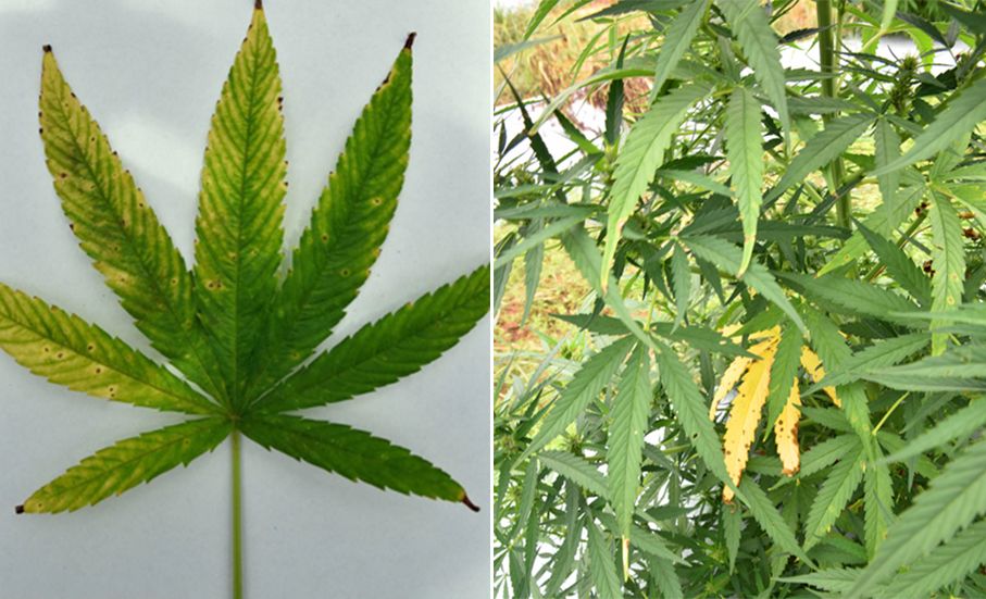
Credit: Rui Yang, Joshua Freeman, and Mathews Paret; UF/IFAS NFREC
Signs & Symptoms
Small necrotic leaf spots with yellow or chlorotic halo, leaf yellowing, and blighting.
Disease Cycle
This bacterial pathogen survives in infected plant material. Like all plant pathogenic bacteria, it enters the plant through natural openings or wounds. Also, the disease intensity can increase with excessive moisture or extended leaf wetness periods. Little information is available about this disease complex on hemp.
Viral Diseases
Signs & Symptoms
Symptoms of viral infections in plants include mosaic, mottling, and yellowing of the leaves. Plant stunting and epinasty (irregular growth) may also occur.
Hemp viral diseases in Florida have not been fully surveyed to understand their causal agents and spread.
Plant Parasitic Nematodes
Nematodes – Meloidogyne spp. and others
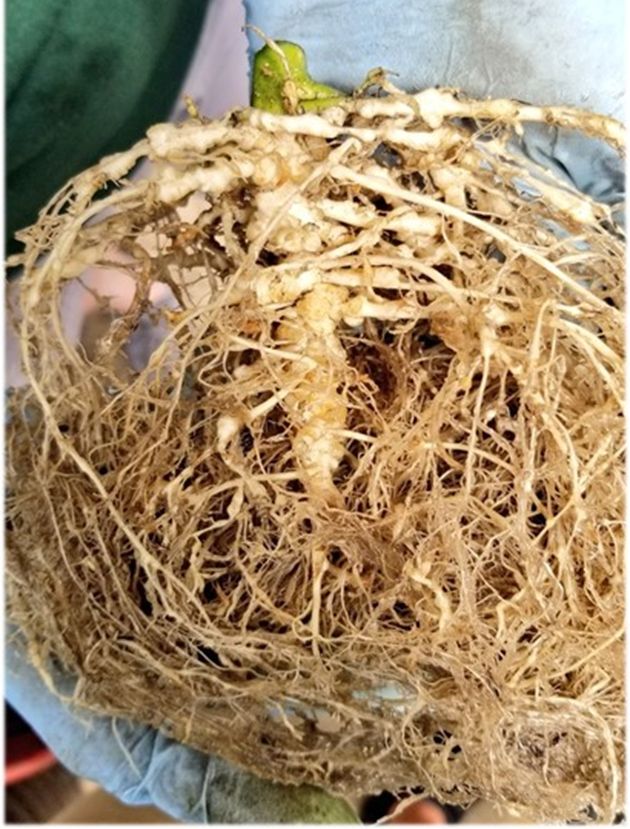
Credit: Jacqueline Coburn and Johan Desaeger, UF/IFAS GCREC
Signs & Symptoms
Symptoms of root-knot nematode (RKN) include yellowing of the canopy, wilting during heat stress, reduced plant vigor, and stunting. Symptoms usually appear in irregular or non-uniform patterns in the field. The symptoms will vary depending on the severity of the infestation and the susceptibility of the host plant. The most prominent symptom is the misshaping of the roots and the formation of galls/knots.
Disease Cycle
Nematodes are microscopic roundworms that typically live in the soil. Plant parasitic nematodes feed on plant roots and affect both water and nutrient uptake. Root-knot nematodes are endoparasites, living inside root galls that they created to facilitate feeding and reproduction. Several species of root-knot RKN are capable of infecting hemp.
Disease Management in Hemp
There is currently limited information on hemp commercial production and a limited number of registered pesticides on hemp in Florida. We recommend implementing an Integrated Pest Management (IPM) approach to manage and minimize the impact of the various potential diseases of hemp. General Core applicable IPM strategies can be found in “Chapter 4. Integrated Pest Management” in the Vegetable Production Handbook of Florida.
Disease Management Resources
(Accessed February 6, 2024)
UF/IFAS Hemp Program & Plant Pathology. https://programs.ifas.ufl.edu/hemp/ & https://plantpath.ifas.ufl.edu/extension/hemp-diseases/
“Chapter 4. Integrated Pest Management,” Vegetable Production Handbook of Florida. https://edis.ifas.ufl.edu/publication/CV298
FDACS Registered List of Pesticides. https://ccmedia.fdacs.gov/content/download/91490/file/fdacs-guide-list-pesticide-products-for-use-on-hemp-2020.pdf
FDACS Hemp and Pesticides. https://ccmedia.fdacs.gov/content/download/91491/file/hemp-pesticides-brochure.pdf
University of Kentucky, Hemp Research, Extension, and Education; Diseases and Insect Pests. https://hemp.ca.uky.edu/diseases-insect-pests
Other Contributor Credits
Reviewers
Philip Harmon, UF/IFAS Department of Plant Pathology
Morgan Pinkerton, UF/IFAS Extension Seminole County
Zachary Brym, UF/IFAS Department of Agronomy
Photo Contributors
Brenda Kennedy, University of Kentucky, Department of Plant Pathology
Christian Christensen, UF/IFAS Extension
Evelyne (Prissy) Fletcher, UF/IFAS Extension
Jacqueline Coburn, UF/IFAS Gulf Coast Research and Education Center
Johan Desaeger, UF/IFAS Gulf Coast Research and Education Center
Joshua Freeman, (formerly) UF/IFAS North Florida Research and Education Center
Katherine E. M. Hendricks, UF/IFAS Extension, Plant Disease Diagnostic Clinic
Marcus Marin, UF/IFAS Gulf Coast Research and Education Center
Mathews Paret, UF/IFAS North Florida Research and Education Center
Nan-Yi Wang, (formerly) UF/IFAS Gulf Coast Research and Education Center
Natalia Peres, UF/IFAS Gulf Coast Research and Education Center
Nicole Gauthier, University of Kentucky, Department of Plant Pathology
Shouhua Wang, Nevada Department of Agriculture
Thomas Isakeit, Texas A&M University
Rui Yang, UF/IFAS North Florida Research and Education Center
Zachariah Hansen, University of Tennessee Extension
References
Beckerman, J., H. Nisonson, N. Albright, and T. Creswell. 2017. “First Report of Pythium aphanidermatum Crown and Root Rot of Industrial Hemp in the United States.” Plant Disease 101 (6): 1038. https://doi.org/10.1094/PDIS-09-16-1249-PDN
Beckerman, J., J. Stone, G. Ruhl, and T. Creswell. 2018. “First Report of Pythium ultimum Crown and Root Rot of Industrial Hemp in the United States.” Plant Disease 102 (10): 2045. https://doi.org/10.1094/PDIS-12-17-1999-PDN
Garfinkel, A. R. 2020. “Three Botrytis Species Found Causing Gray Mold on Industrial Hemp (Cannabis sativa) in Oregon.” Plant Disease 104 (7): 2026. https://doi.org/10.1094/PDIS-01-20-0055-PDN
Gauthier, N., M. Rahnama, and K. Leonbergerr. 2021. “Septoria Leaf Spot of Field Hemp.” PPS-AG-H-01. University of Kentucky, Cooperative Extension Service. https://plantpathology.ca.uky.edu/files/ppfs-ag-h-01.pdf
Giles, G., E. J. Indermaur, J. L. Gonzalez-Giron, T. Q. Hermann, S. Shelnutt, J. Starr, K. Myers, et al. 2023. “First Report of Downy Mildew caused by Pseudoperonospora cannabina on Cannabis sativa in New York.” Plant Disease 107 (5): 1638. https://doi.org/10.1094/PDIS-08-22-1930-PDN
Gwinn, K. D., Z. Hansen, H. Kelly, and B. H. Ownley. 2022. “Diseases of Cannabis sativa Caused by Diverse Fusarium Species.” Frontiers in Agronomy 3. https://doi.org/10.3389/fagro.2021.796062
Hu, J. 2021. “First Report of Crown and Root Rot Caused by Pythium myriotylum on Hemp (Cannabis sativa) in Arizona.” Plant Disease 105 (9): 2736. https://doi.org/10.1094/PDIS-12-20-2712-PDN
Iriarte, F. B., J. Freeman, R. Yang, and M. Paret. 2021. “Industrial Hemp Pilot Program 2019-2020: Hemp Diseases in North Florida.” PowerPoint presentation, UF/IFAS North Florida Research and Education Center. https://plantpath.ifas.ufl.edu/u-scout/ewExternalFiles/HEMP-DISEASES-FL_Iriarte_Paret.pdf
Marin, M. V., J. Coburn, J. Desaeger, and N. A. Peres. 2020. “First Report of Cercospora Leaf Spot Caused by Cercospora cf. flafellaris on Industrial Hemp in Florida.” Plant Disease 104 (5): 1536. https://doi.org/10.1094/PDIS-11-19-2287-PDN
Marin, M. V., N. -Y. Wang, J. Coburn, J. Desaeger, and N. A. Peres. 2020. “First Report of Curvularia pseudobrachyspora Causing Leaf Spot on Hemp (Cannabis sativa) in Florida.” Plant Disease 104 (12): 3262. https://doi.org/10.1094/PDIS-03-20-0546-PDN
Marin, M. V., N. -Y. Wang, J. Coburn, J. Desaeger, and N. A. Peres. 2021. “First Report of Diaporthe phaseolorum Causing Stem Canker of Hemp (Cannabis sativa).” Plant Disease 105 (7): 2018. https://doi.org/10.1094/PDIS-06-20-1174-PDN
McGehee, C. S., P. Apicella, R. Raudales, G. Berkowitz, Y. Ma, S. Durocher, and J. Lubell. 2019. “First Report of Root Rot and Wilt Caused by Pythium myriotylum on Hemp (Cannabis sativa) in the United States.” Plant Disease 103 (12): 3288. https://doi.org/10.1094/PDIS-11-18-2028-PDN
Pane, A., L. S. Cosentino, V. Copani, and S. O. Cacciola. 2007. “First Report of Southern Blight Caused by Sclerotium rolfsii on Hemp (Cannabis sativa) in Sicily and Southern Italy.” Plant Disease 91 (5): 636. https://doi.org/10.1094/PDIS-91-5-0636A
Pfeufer, E., C. Bradley, and N. Gautheir. 2018. “Southern Blight.” PPFS-GEN-16. University of Kentucky, Cooperative Extension Service. https://plantpathology.ca.uky.edu/files/ppfs-gen-16.pdf
Szarka, D., B. Amsden, J. Beale, E. Dixon, C. L. Schardl, and N. Gauthier. 2020. “First Report of Hemp Leaf Spot Caused by a Bipolaris Species on Hemp (Cannabis sativa) in Kentucky.” Plant Health Progress 21 (2): 82–84. https://doi.org/10.1094/PHP-01-20-0004-BR
Szarka D., L. Tymon, B. Amsden, E. Dixon, J. Judy, and N. Gauthier. 2019. “First Report of Powdery Mildew Caused by Golovinomyces spadiceus on Industrial Hemp (Cannabis Sativa) in Kentucky.” Plant Disease 103 (7): 1773. https://doi.org/10.1094/PDIS-01-19-0049-PDN